Copyright
©The Author(s) 2019.
World J Clin Cases. Nov 26, 2019; 7(22): 3772-3777
Published online Nov 26, 2019. doi: 10.12998/wjcc.v7.i22.3772
Published online Nov 26, 2019. doi: 10.12998/wjcc.v7.i22.3772
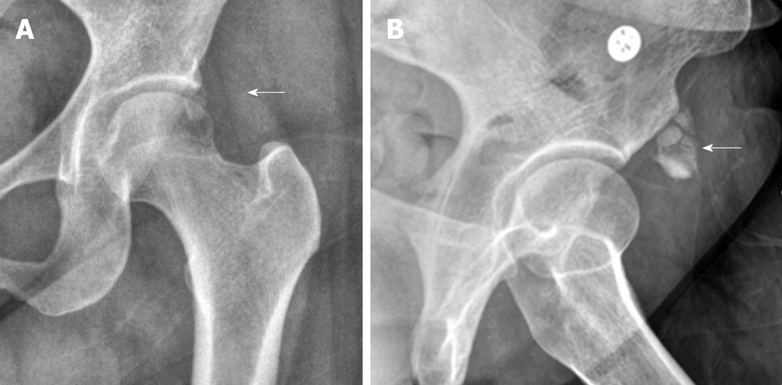
Figure 1 An anteroposterior radiography of the left hip.
A: An anteroposterior radiography of the left hip showing an amorphous calcification (white arrow) adjacent to the anterior inferior iliac spine which is the attachment site of the rectus tendon, suggesting calcific tendinopathy of rectus femoris; B: The lesion (white arrow) was more clearly defined at the lateral view.
Figure 2 Longitudinal ultrasonographic image.
A: Longitudinal ultrasonographic image showing a solid calcific nodule (white arrow) at 1.70 cm × 0.85 cm in size adjacent to the insertion site of the rectus femoris tendon; B: An arc shaped lesion (white arrow) of 1.43 cm × 0.94 cm was also defined at the transverse ultrasonographic image. AIIS: Anterior inferior iliac spine; RF: Rectus femoris.
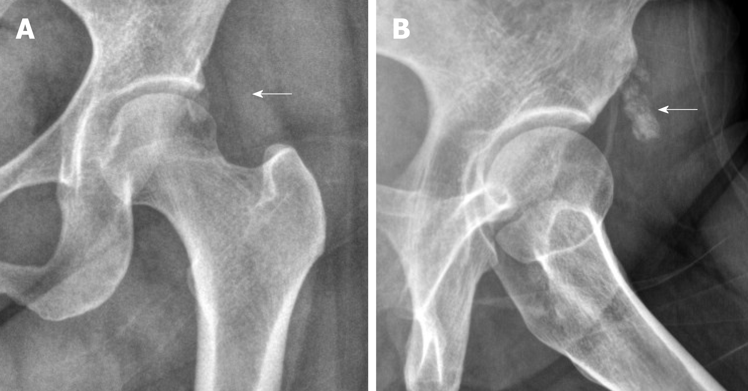
Figure 3 Simple radiography.
A: The size of calcification was almost not reduced in simple radiography at the final follow-up; B: The lesion (white arrow) was still clearly defined at lateral view.
Figure 4 Ultrasonographic image of rectus femoris calcification after extracorporeal shock wave therapy.
A: The calcium deposit was decreased (1.42 cm × 0.61 cm) on longitudinal image; B: Huge calcification disintegrates into smaller pieces on transverse image. AIIS: Anterior inferior iliac spine; RF: Rectus femoris.
- Citation: Lee CH, Oh MK, Yoo JI. Ultrasonographic evaluation of the effect of extracorporeal shock wave therapy on calcific tendinopathy of the rectus femoris tendon: A case report. World J Clin Cases 2019; 7(22): 3772-3777
- URL: https://www.wjgnet.com/2307-8960/full/v7/i22/3772.htm
- DOI: https://dx.doi.org/10.12998/wjcc.v7.i22.3772


