Copyright
©The Author(s) 2019.
World J Clin Cases. Oct 26, 2019; 7(20): 3372-3376
Published online Oct 26, 2019. doi: 10.12998/wjcc.v7.i20.3372
Published online Oct 26, 2019. doi: 10.12998/wjcc.v7.i20.3372
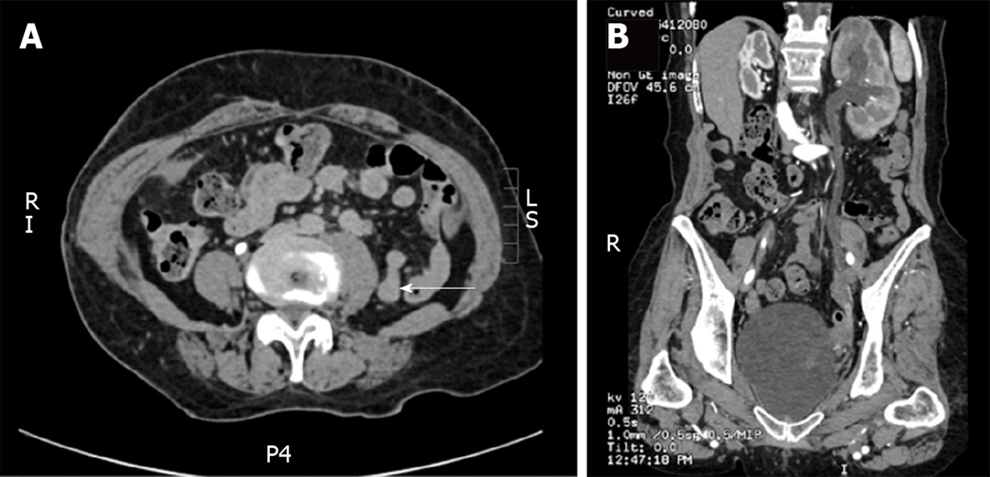
Figure 1 Preoperative computed tomography scanning.
A: Coronal plane; B: Sagittal plane. Preoperative computed tomography scans of the urinary system in the coronal plane and sagittal plane views revealing the left ureteral mass and left ureteral dilatation.
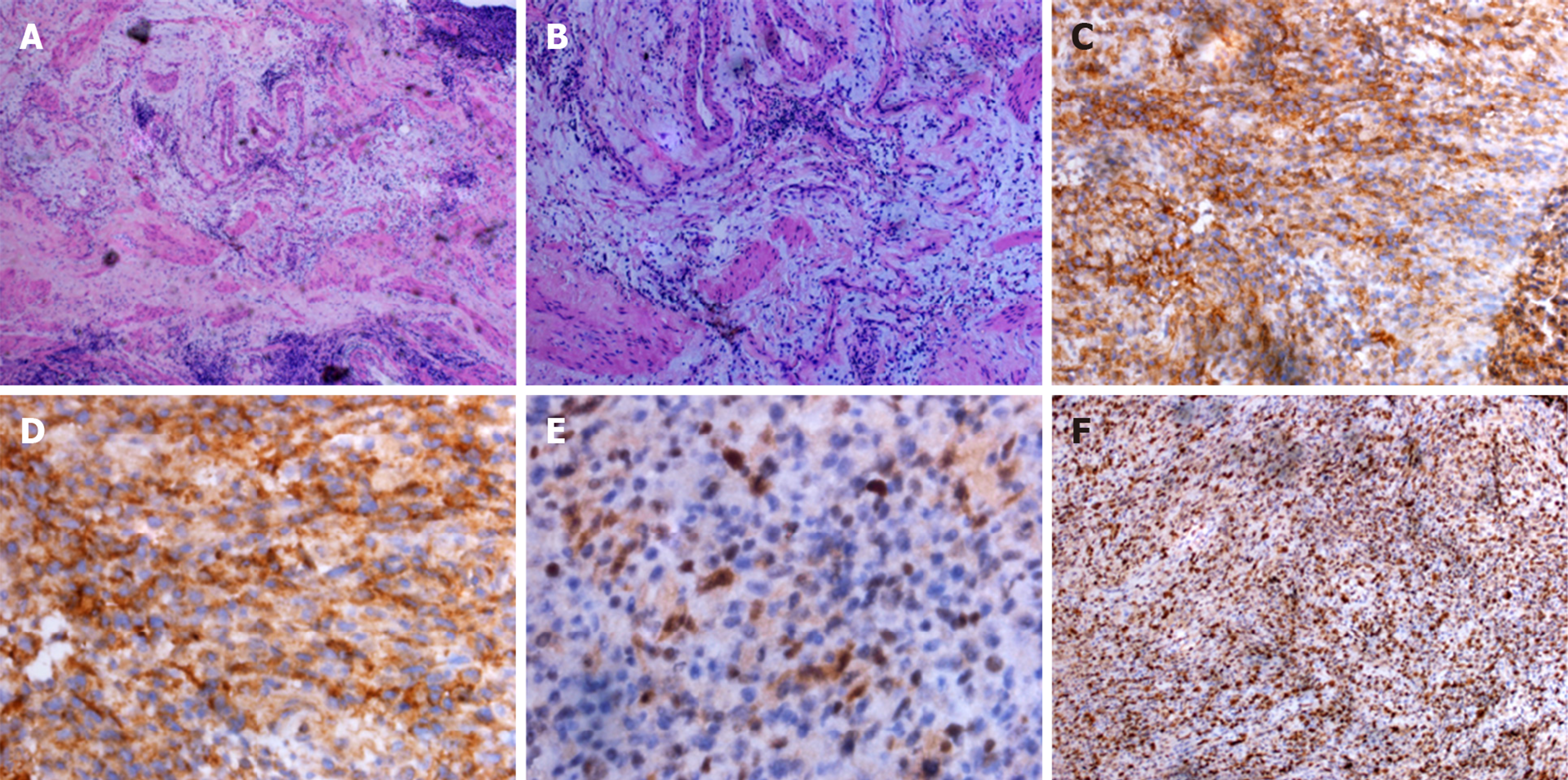
Figure 2 Microscopic and pathologic images.
A, B: Microscopic view of the left ureter tumor stained with hematoxylin and eosin. Histopathologic features suggested that it was a small cell malignant tumor of the left ureter; C-F: Immunohistochemical staining was positive for CD99 (C, D), transducin-like enhancer protein 1 (E), and Ki67 (F).
- Citation: Li XX, Bi JB. Ureteral Ewing’s sarcoma in an elderly woman: A case report. World J Clin Cases 2019; 7(20): 3372-3376
- URL: https://www.wjgnet.com/2307-8960/full/v7/i20/3372.htm
- DOI: https://dx.doi.org/10.12998/wjcc.v7.i20.3372


