Copyright
©The Author(s) 2019.
World J Clin Cases. Sep 26, 2019; 7(18): 2815-2822
Published online Sep 26, 2019. doi: 10.12998/wjcc.v7.i18.2815
Published online Sep 26, 2019. doi: 10.12998/wjcc.v7.i18.2815
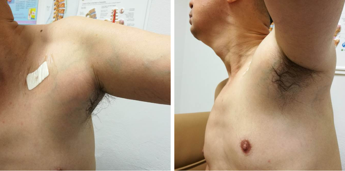
Figure 1 Clinical photos showing a large lump in the left axillary region of a 48-year-old Thai male patient.
Figure 2 Histologic images.
A, B: Histologic images of tumor tissue demonstrating sheets and cords (A) of relatively uniform tumor cells with foamy cytoplasm and round to oval hyperchromatic nuclei without atypia (B); C: The mitotic count was 0 per 50 high power field. The Ki-67 was 0-5%.
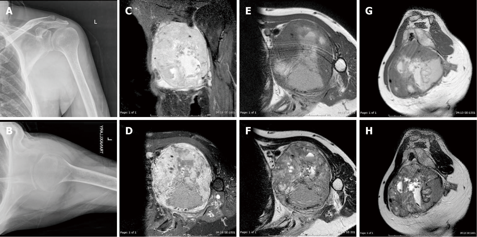
Figure 3 Radiographs and preoperative magnetic resonance imaging.
A, B: Preoperative plain coronal (A) and trans-scapular (B) radiographs of the left shoulder showing a large soft tissue shadow in the axilla; C-H: Preoperative magnetic resonance imaging: coronal (C) and transverse (D) fat suppressed T1-weighted images after intravenous administration of gadolinium, transverse T1-weighted (E), transverse T2-weighted (F), sagittal T1-weighted (G), and sagittal T2-weighted (H) images demonstrating a large heterogeneous, contrast-enhancing mass bulging from the posterior cord of the brachial plexus, encasing the axillary artery and vein, and occupying the entire axillary space.
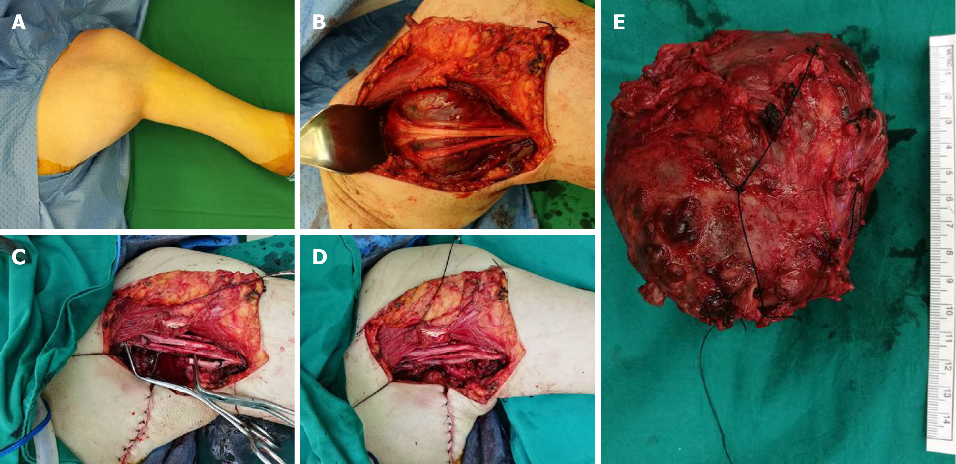
Figure 4 Intraoperative pictures.
A: Intraoperative pictures demonstrating a large mass in the left axilla; B: The tumor arose within the brachial plexus and was severely adherent to the left axillary artery and posterior cord; C, D: Complete surgical resection of the tumor along with the abutted segment of the axillary artery was performed (C), followed by saphenous vein graft reconstruction (D); E: Representative picture of the gross tumor showing a large red-brown multinodular mass associated with a segment of the axillary artery.
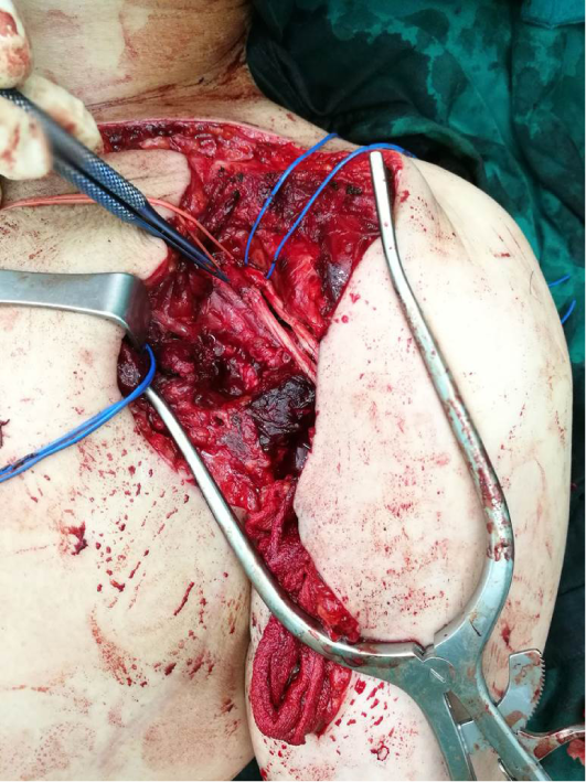
Figure 5 The second stage sural nerve grafting to the radial and the axillary nerve was performed to treat postoperative incomplete brachial plexopathy, without any sign of re-innervation at 6-mo follow-up.
- Citation: Thanindratarn P, Chobpenthai T, Phorkhar T, Nelson SD. Glomus tumor of uncertain malignant potential of the brachial plexus: A case report. World J Clin Cases 2019; 7(18): 2815-2822
- URL: https://www.wjgnet.com/2307-8960/full/v7/i18/2815.htm
- DOI: https://dx.doi.org/10.12998/wjcc.v7.i18.2815


