Copyright
©The Author(s) 2019.
World J Clin Cases. Aug 26, 2019; 7(16): 2360-2366
Published online Aug 26, 2019. doi: 10.12998/wjcc.v7.i16.2360
Published online Aug 26, 2019. doi: 10.12998/wjcc.v7.i16.2360
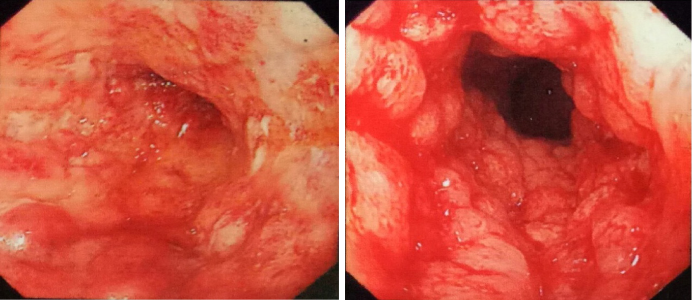
Figure 1 Endoscopy demonstrated total colonic wall thickening, erosions in luminal surface of colon, hyperemia, friability, bleeding and ulcerations.
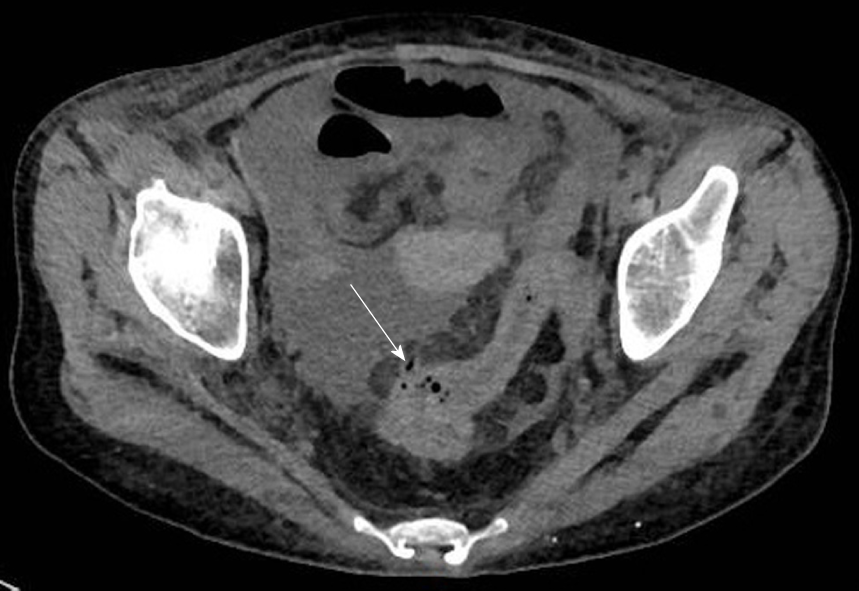
Figure 2 Thin flaws and perforations at rectosigmoid colon and a large amount of fluid in abdominal cavity and pelvis were found by emergency computed tomography.
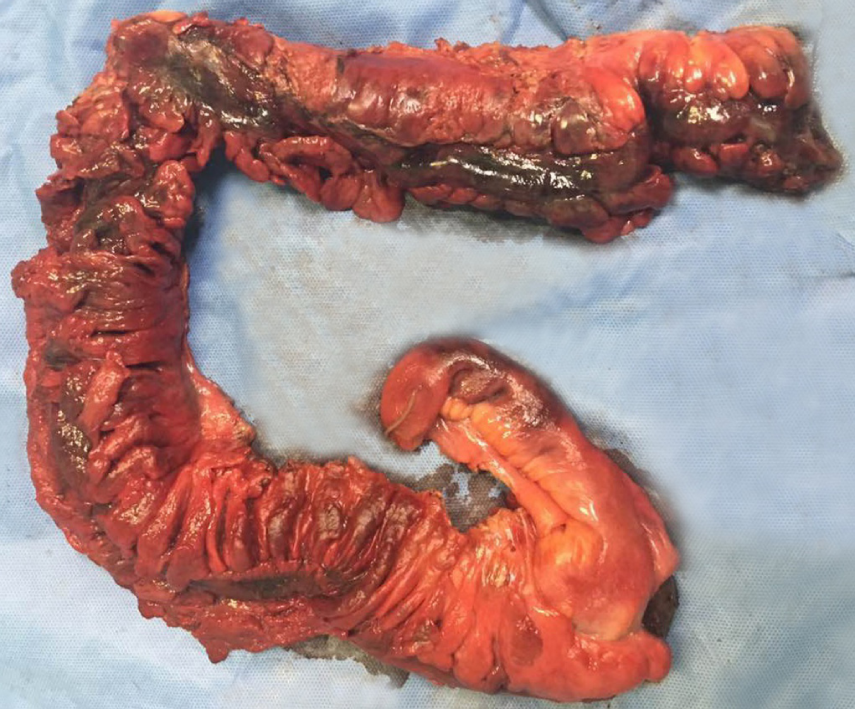
Figure 3 Total colonic necrosis was seen during operation.
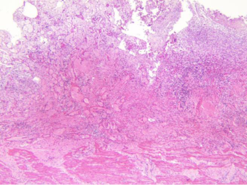
Figure 4 Histopathological examination of the resected colon in the patient showed extensive hemorrhage, necrosis and exudation involving the whole intestinal wall and the extraserous adipose tissue.
Figure 5 Plain computed tomography scanning and contrast enhancement of abdomen display thrombosis in the trunk of portal vein and its intrahepatic branches, the superior mesenteric vein and the splenic vein.
- Citation: Zhu MY, Sun LQ. Ulcerative colitis complicated with colonic necrosis, septic shock and venous thromboembolism: A case report. World J Clin Cases 2019; 7(16): 2360-2366
- URL: https://www.wjgnet.com/2307-8960/full/v7/i16/2360.htm
- DOI: https://dx.doi.org/10.12998/wjcc.v7.i16.2360


