Copyright
©The Author(s) 2019.
World J Clin Cases. Aug 6, 2019; 7(15): 2075-2080
Published online Aug 6, 2019. doi: 10.12998/wjcc.v7.i15.2075
Published online Aug 6, 2019. doi: 10.12998/wjcc.v7.i15.2075
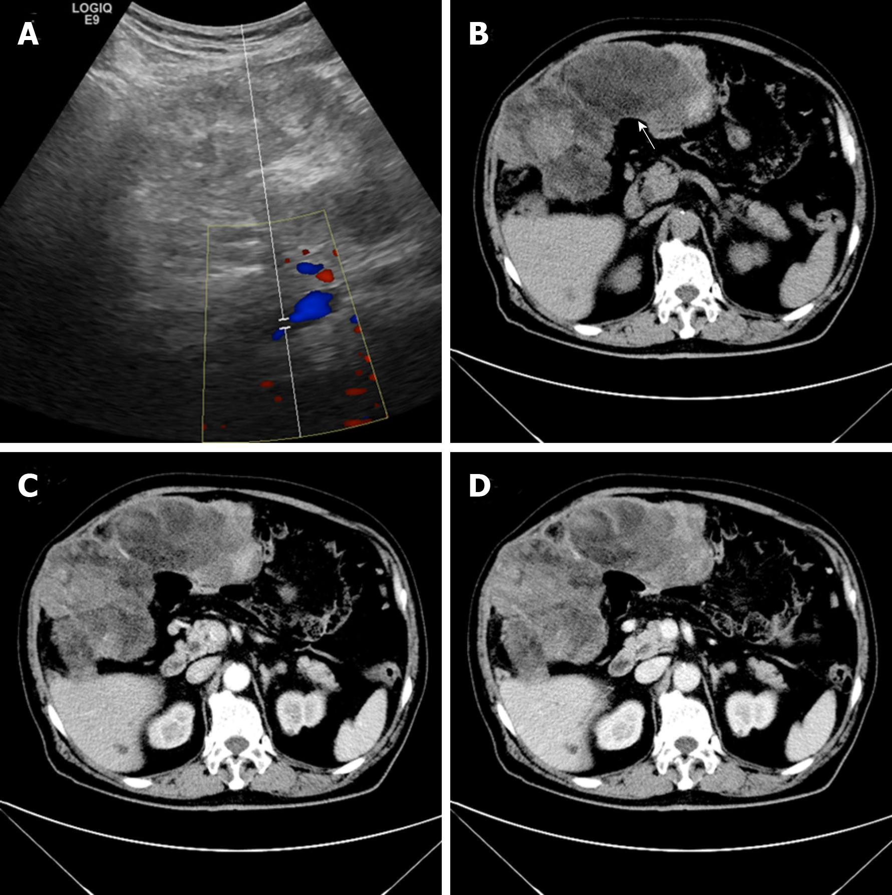
Figure 1 Imaging diagnosis.
A: Color Doppler image showing blood flow; B: Computed tomography with contrast (white arrow, fat density); C: Uneven heterogeneous enhancement in the arterial phase; D: Delayed enhancement in the venous phase.
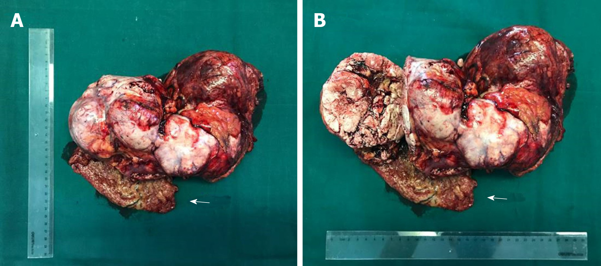
Figure 2 The 30 cm × 15 cm × 8 cm heterogeneous tumor with necrosis (A, B).
Arrows invaded abdominal wall tissue.
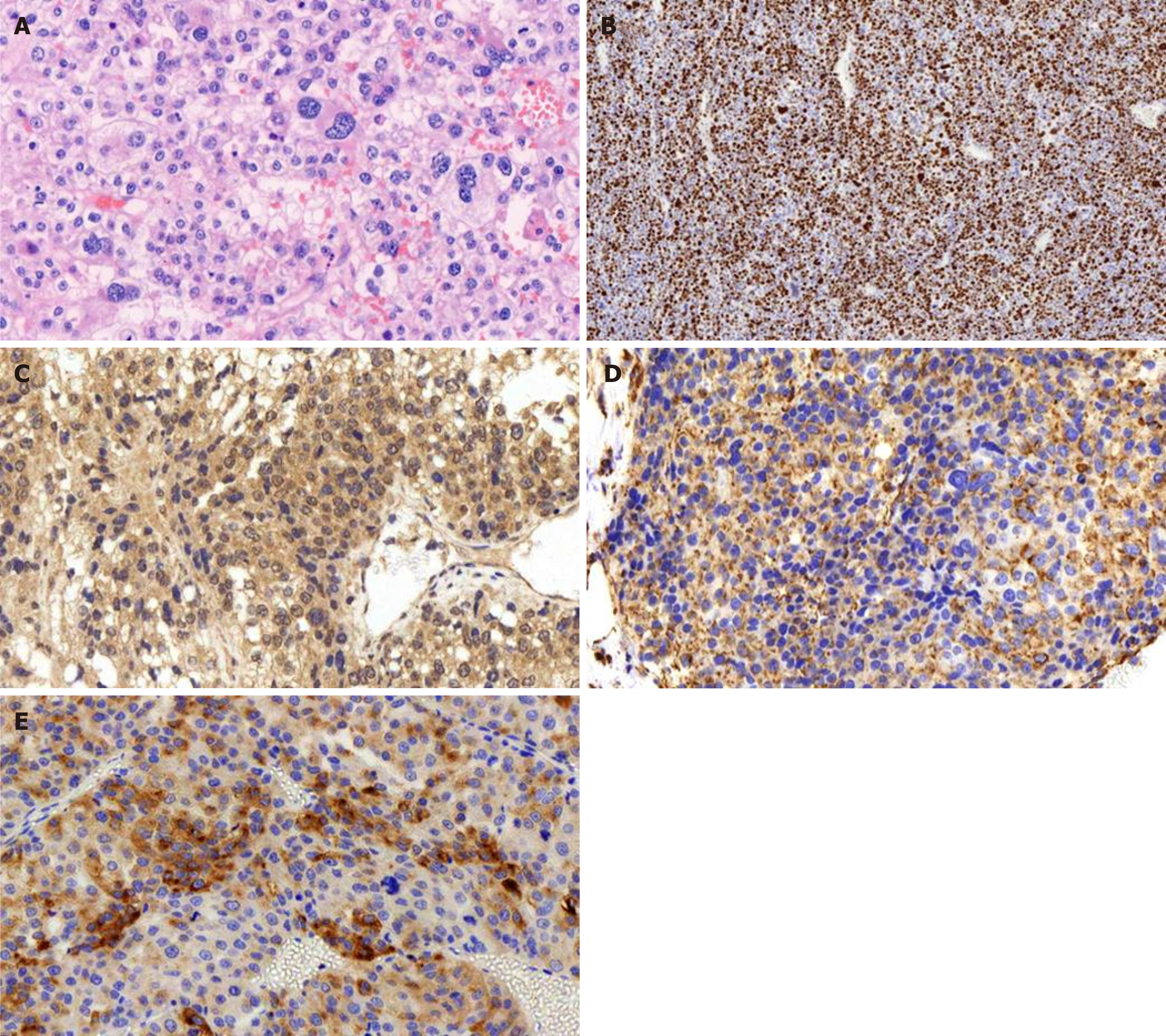
Figure 3 Histological staining.
A: Histological staining showing significant nuclear atypia, necrosis, and pleomorphism of the tumor cells (magnification, 200×); B: The Ki-67 labeling index was > 80% (IHC staining; magnification, 50×); C-D: Positive IHC staining for (C) melan-A, (D) vimentin and (E) synaptophysin (magnification, 200×).
- Citation: Zhou DK, Liu ZH, Gao BQ, Wang WL. Giant nonfunctional ectopic adrenocortical carcinoma on the anterior abdominal wall: A case report. World J Clin Cases 2019; 7(15): 2075-2080
- URL: https://www.wjgnet.com/2307-8960/full/v7/i15/2075.htm
- DOI: https://dx.doi.org/10.12998/wjcc.v7.i15.2075


