Copyright
©The Author(s) 2019.
World J Clin Cases. Jul 6, 2019; 7(13): 1696-1702
Published online Jul 6, 2019. doi: 10.12998/wjcc.v7.i13.1696
Published online Jul 6, 2019. doi: 10.12998/wjcc.v7.i13.1696
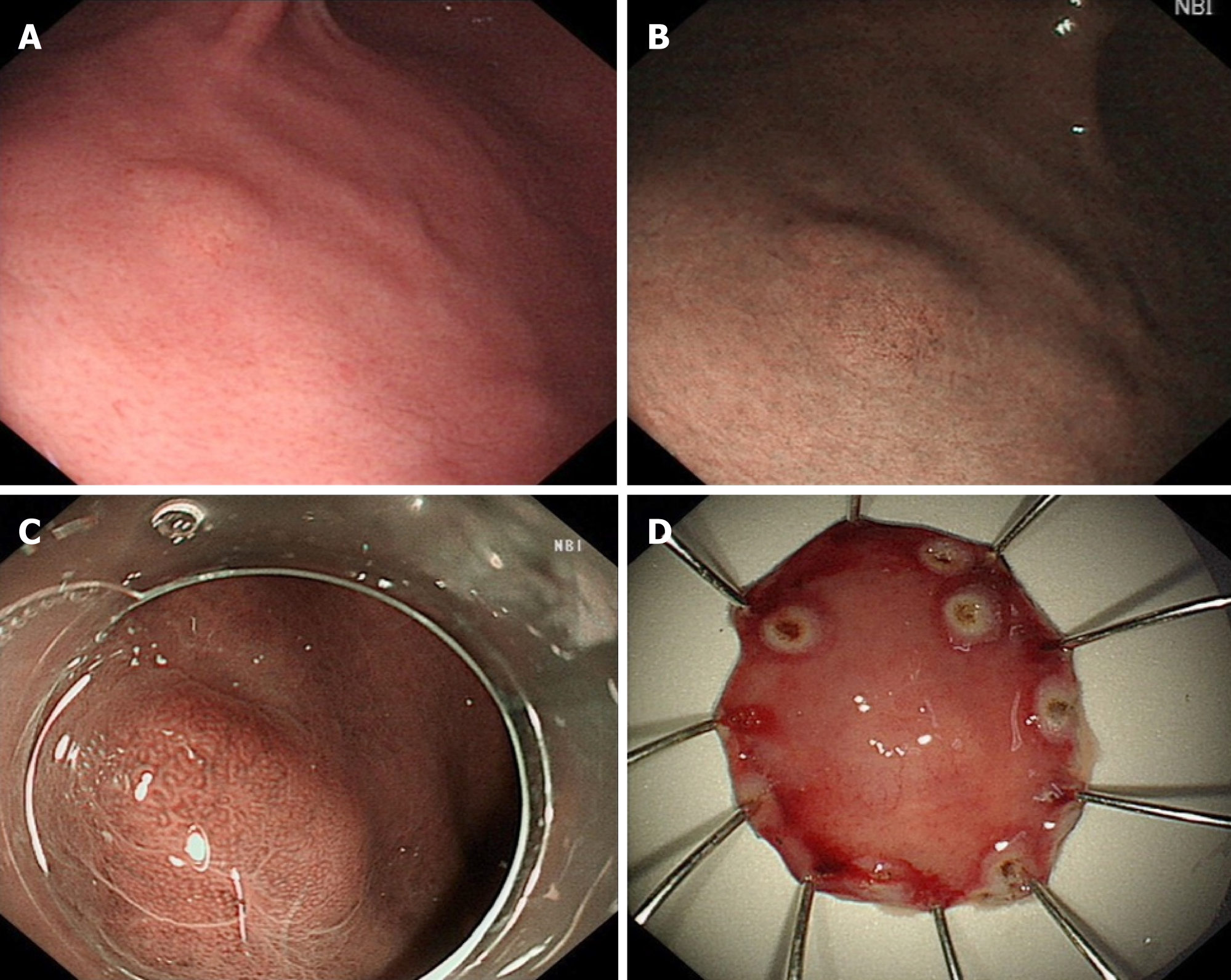
Figure 1 Endoscopic findings.
A, B: A yellowish submucosal tumor-like elevated lesion is observed with white light endoscopy (A) and narrowband imaging (B) in the anterior wall of the lower third of the gastric body; C: It showed an enlarged microvascular pattern and a mildly disordered microsurface pattern especially prominent in the upper part of the lesion; D: On assessment of the endoscopic submucosal dissection specimen, a tumor measuring 5 mm × 6 mm was identified in the submucosal layer.
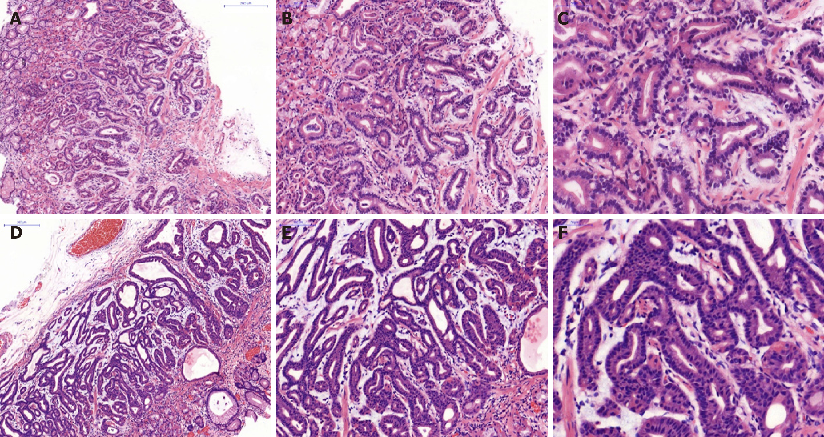
Figure 2 Histopathological findings (hematoxylin and eosin staining).
A-F: Well-differentiated adenocarcinoma with numerous fundic gland-like cells were observed in the biopsy (A, B, and C) and endoscopic submucosal dissection specimen (D, E and F), which were crowded and disordered in a layer of the lamina propria mucosae, and formed the so-called “endless glands” pattern. Magnification: A and D: 100 ×; B and E: 200 ×; C and F: 400 ×.
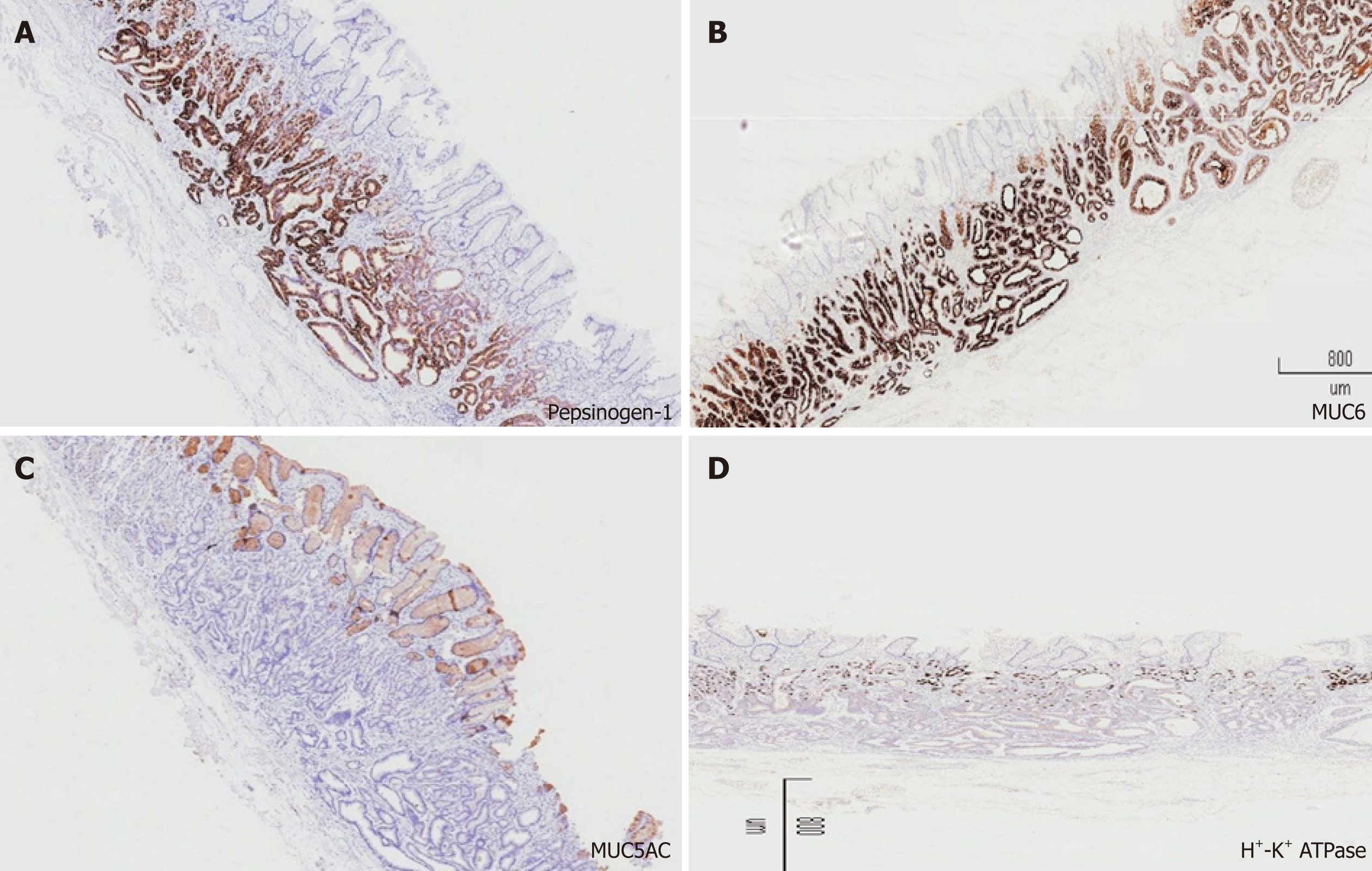
Figure 3 Immunohistochemical findings.
Magnification of all photographs is 40 ×. A-D: The tumor cells of gastric adenocarcinoma of fundic gland type were strongly and diffusely reactive for pepsinogen-I (A) and MUC6 (B), negative for MUC5AC (C) and H+/K+-ATPase (D). MUC6: Mucin 6.
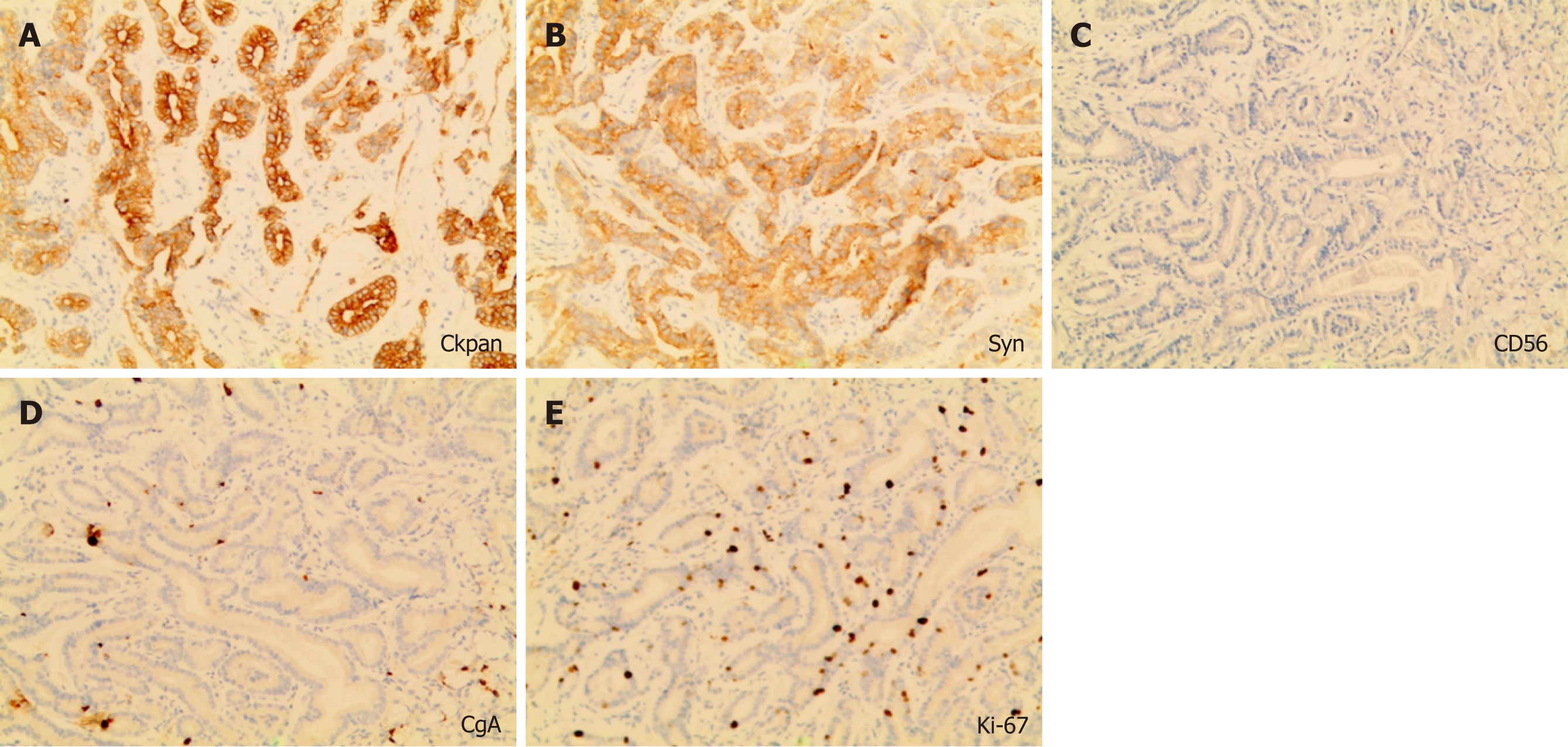
Figure 4 Immunohistochemical findings.
A, B: Magnification of all photographs is 200 ×. The tumor was strongly positive for Ckpan (A), scattered positivity for Syn (B); C, D, E: Staining for CgA (C) and CD56 was negative (D), and the Ki-67 labeling index was 4% (E).
- Citation: Yu YN, Yin XY, Sun Q, Liu H, Zhang Q, Chen YQ, Zhao QX, Tian ZB. Gastric adenocarcinoma of fundic gland type after Helicobacter pylori eradication: A case report. World J Clin Cases 2019; 7(13): 1696-1702
- URL: https://www.wjgnet.com/2307-8960/full/v7/i13/1696.htm
- DOI: https://dx.doi.org/10.12998/wjcc.v7.i13.1696


