Copyright
©The Author(s) 2019.
World J Clin Cases. May 26, 2019; 7(10): 1230-1233
Published online May 26, 2019. doi: 10.12998/wjcc.v7.i10.1230
Published online May 26, 2019. doi: 10.12998/wjcc.v7.i10.1230
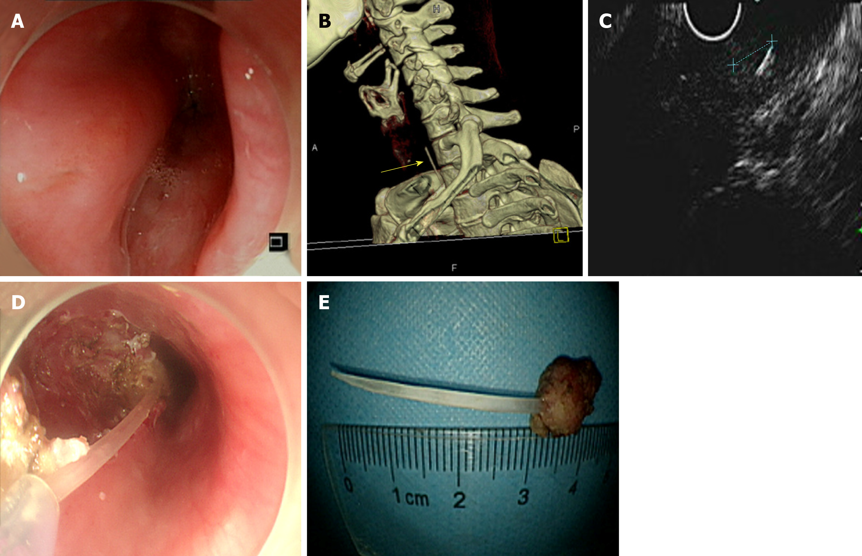
Figure 1 Fish bone removal by endoscopy.
A: Gastroscopic examination suggested that the upper esophageal segment exhibited a strip-shaped submucous bulge; B: Computed tomography suggested that the upper esophageal segment contained a high-density band in the right wall; C: Endoscopic ultrasonography revealed that the upper submucosa of the esophagus exhibited a striped hyperechoic mass; D: Removal of the fish bone by endoscopic submucosal dissection (ESD); E: The fish bone removed by ESD.
- Citation: Wang XM, Yu S, Chen X. Successful endoscopic extraction of a proximal esophageal foreign body following accurate localization using endoscopic ultrasound: A case report. World J Clin Cases 2019; 7(10): 1230-1233
- URL: https://www.wjgnet.com/2307-8960/full/v7/i10/1230.htm
- DOI: https://dx.doi.org/10.12998/wjcc.v7.i10.1230


