Copyright
©The Author(s) 2019.
World J Clin Cases. May 26, 2019; 7(10): 1221-1229
Published online May 26, 2019. doi: 10.12998/wjcc.v7.i10.1221
Published online May 26, 2019. doi: 10.12998/wjcc.v7.i10.1221
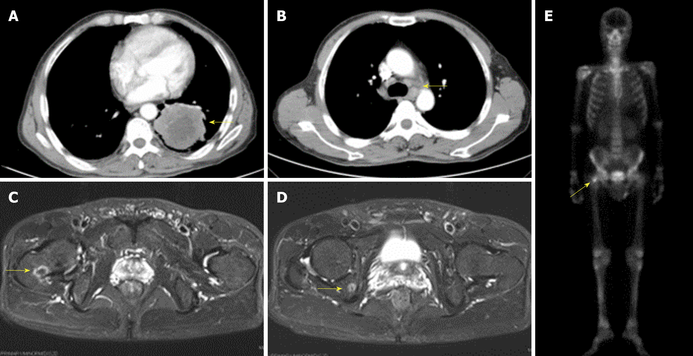
Figure 1 Baseline imaging examinations.
Primary cancer in the inferior lobe of the left lung (A, arrow) with metastases to the hilar and mediastinal lymph nodes (B, arrow) and multiple bones (C-E, arrows).
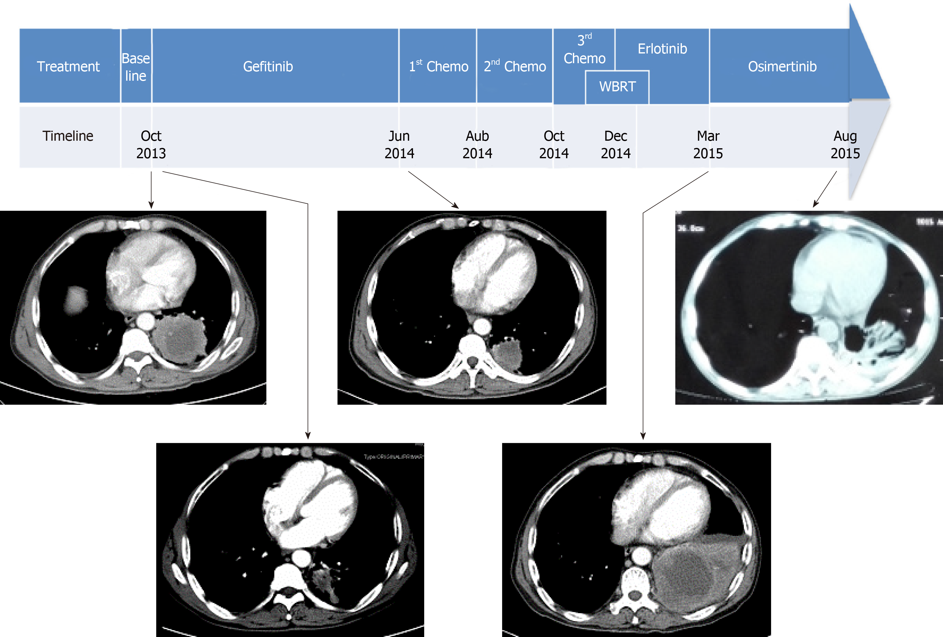
Figure 2 Sequence of anti-cancer treatments across the timeline and imaging evaluation of respective treatment.
The primary tumor had a partial response to treatment with osimertinib. WBRT: Whole brain radiotherapy.
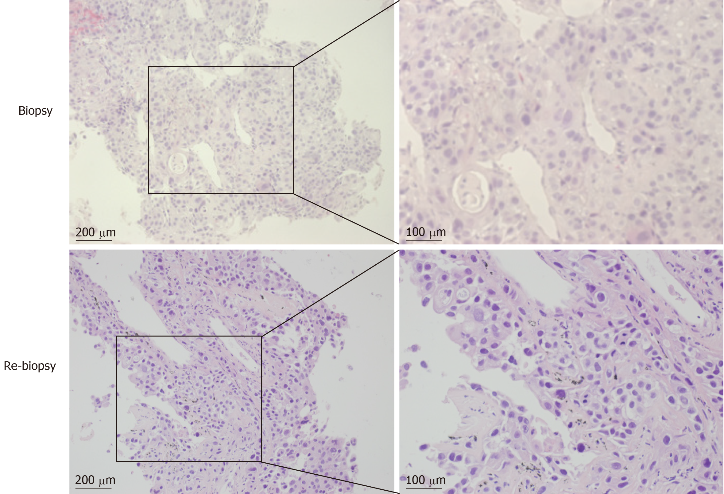
Figure 3 HE staining of specimens from two biopsies (baseline and re-biopsy before osimertinib).
Squamous cell carcinoma was diagnosed by both pathological tests.
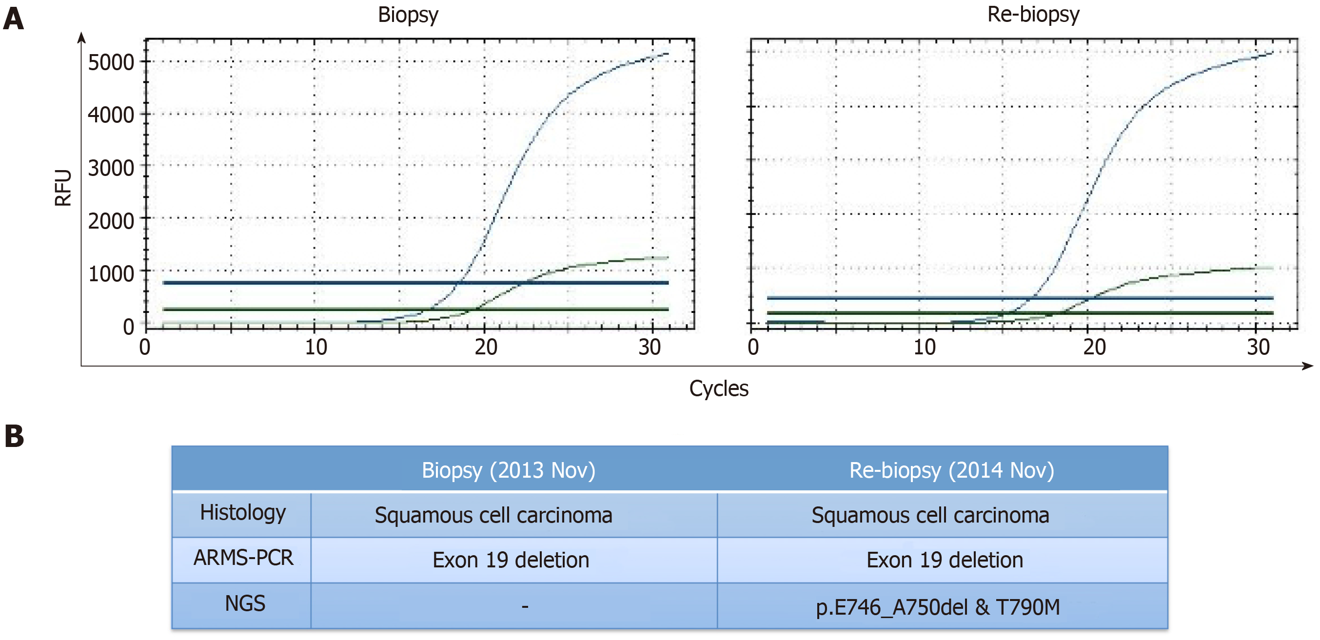
Figure 4 Pathological and gene alteration analyses of the two biopsies.
Amplification refractory mutation system-polymerase chain reaction test only detected exon 19 deletion in both samples (A), whereas next-generation sequencing detected the presence of an EGFR T790M mutation in addition to the exon 19-deletion mutation (p.E746_A750del) of the re-biopsy sample (B). NGS: Next-generation sequencing; ARMS-PCR: Amplification refractory mutation system-polymerase chain reaction.
- Citation: Zhang Y, Chen HM, Liu YM, Peng F, Yu M, Wang WY, Xu H, Wang YS, Lu Y. Significant benefits of osimertinib in treating acquired resistance to first-generation EGFR-TKIs in lung squamous cell cancer: A case report. World J Clin Cases 2019; 7(10): 1221-1229
- URL: https://www.wjgnet.com/2307-8960/full/v7/i10/1221.htm
- DOI: https://dx.doi.org/10.12998/wjcc.v7.i10.1221


