Copyright
©The Author(s) 2019.
World J Clin Cases. May 26, 2019; 7(10): 1191-1199
Published online May 26, 2019. doi: 10.12998/wjcc.v7.i10.1191
Published online May 26, 2019. doi: 10.12998/wjcc.v7.i10.1191
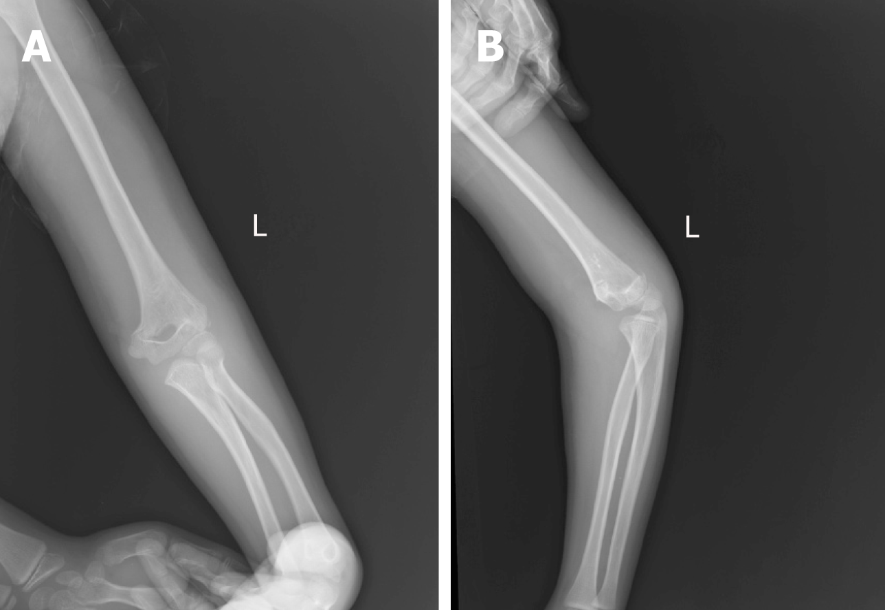
Figure 1 Radiography at the time of injury.
A: Frontal x-ray of the left elbow; B: Lateral x-ray of the left elbow.
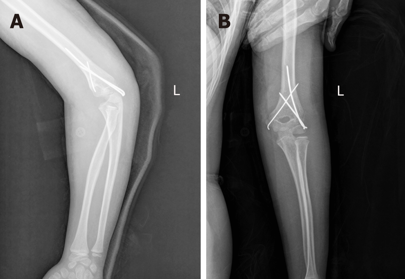
Figure 2 Radiography after the first operation.
A: Lateral s-ray of the left elbow; B: TFrontal x-ray of the left elbow.
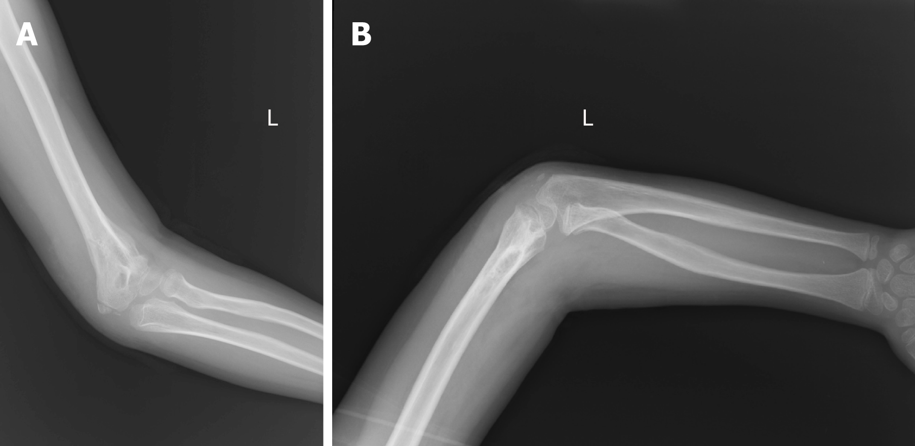
Figure 3 Radiography after removal of the Kirschner wire fixation.
A: Frontal x-ray of the left elbow; B: Lateral x-ray of the left elbow.
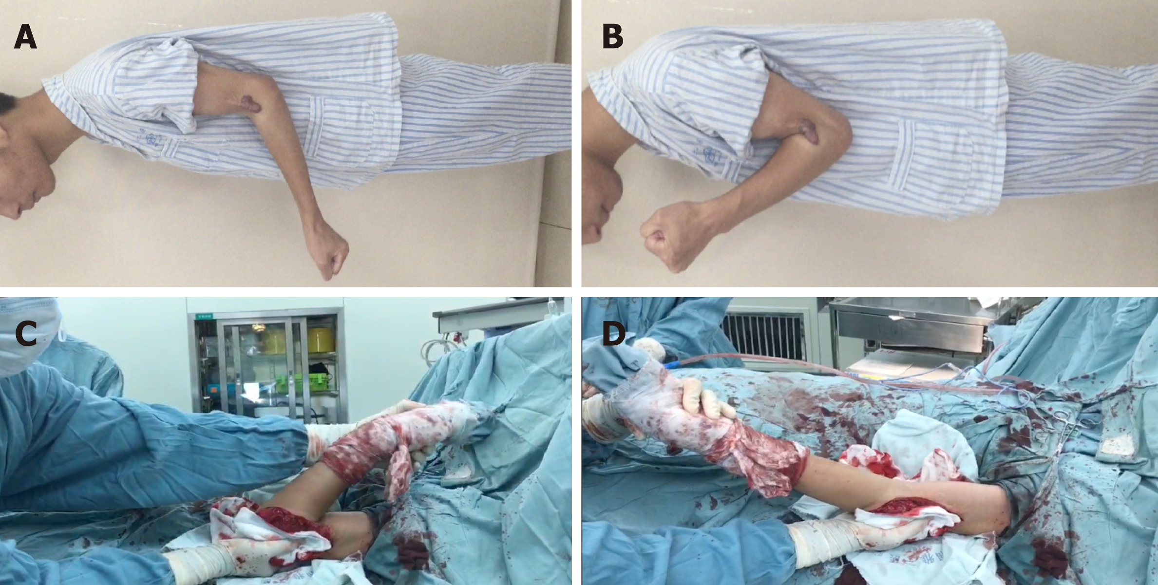
Figure 4 Pre-operative and postoperative flexion/extension range of motion of the left elbow.
A: The best position of extension pre-operatively; B: The best position of flexion pre-operatively; C: The best position of flexion postoperatively; D: The best position of extension postoperatively.
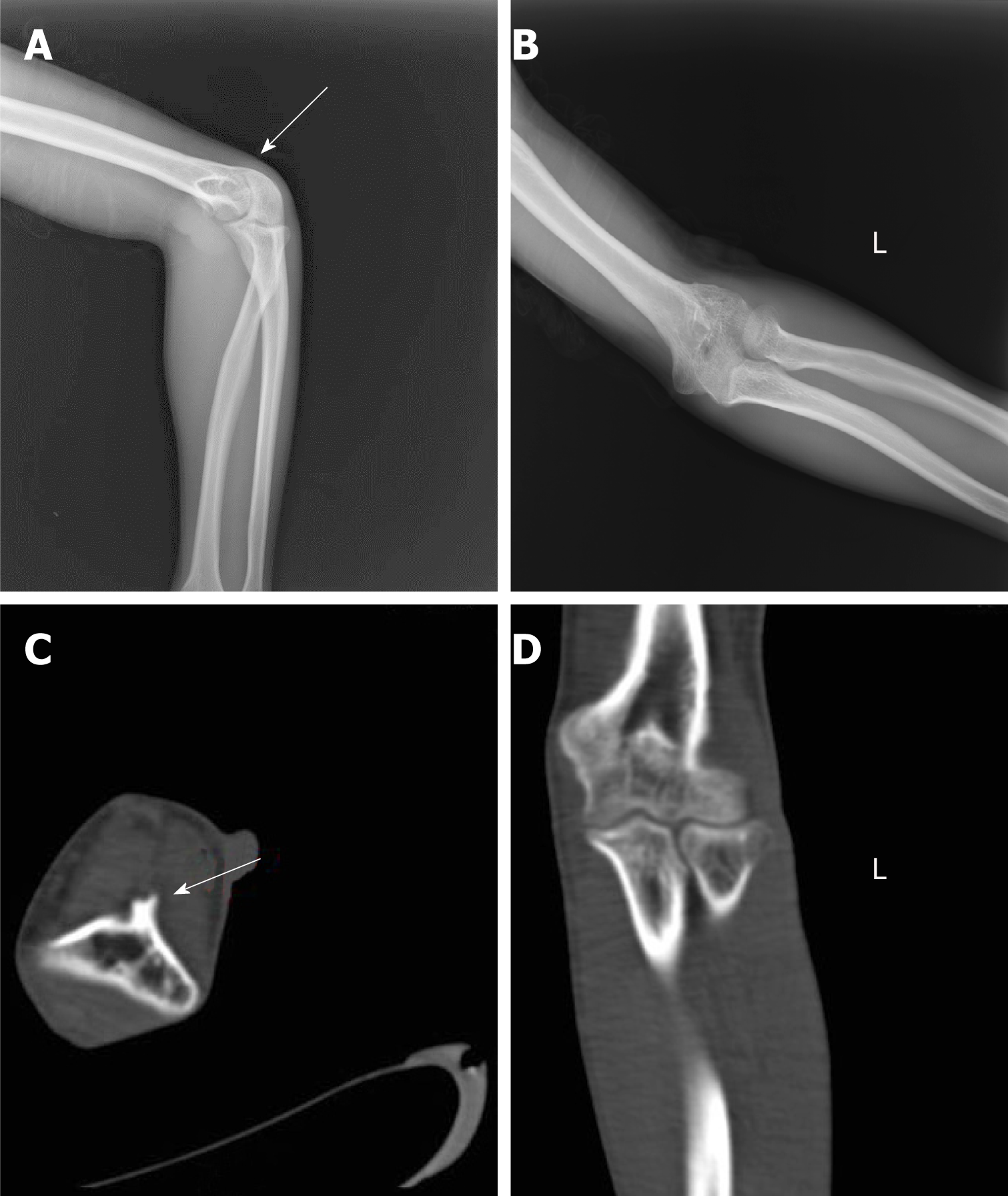
Figure 5 Pre-operative radiograph and representative axillary computed tomography image.
A: Frontal x-ray of the left elbow, with the arrow pointing to the bone connection; B: Lateral x-ray of the left elbow; C: CT scan of the cross section; D: CT coronal scan, with the arrow pointing to the bony block of the humerus coronoid fossa. CT: Computed tomography.
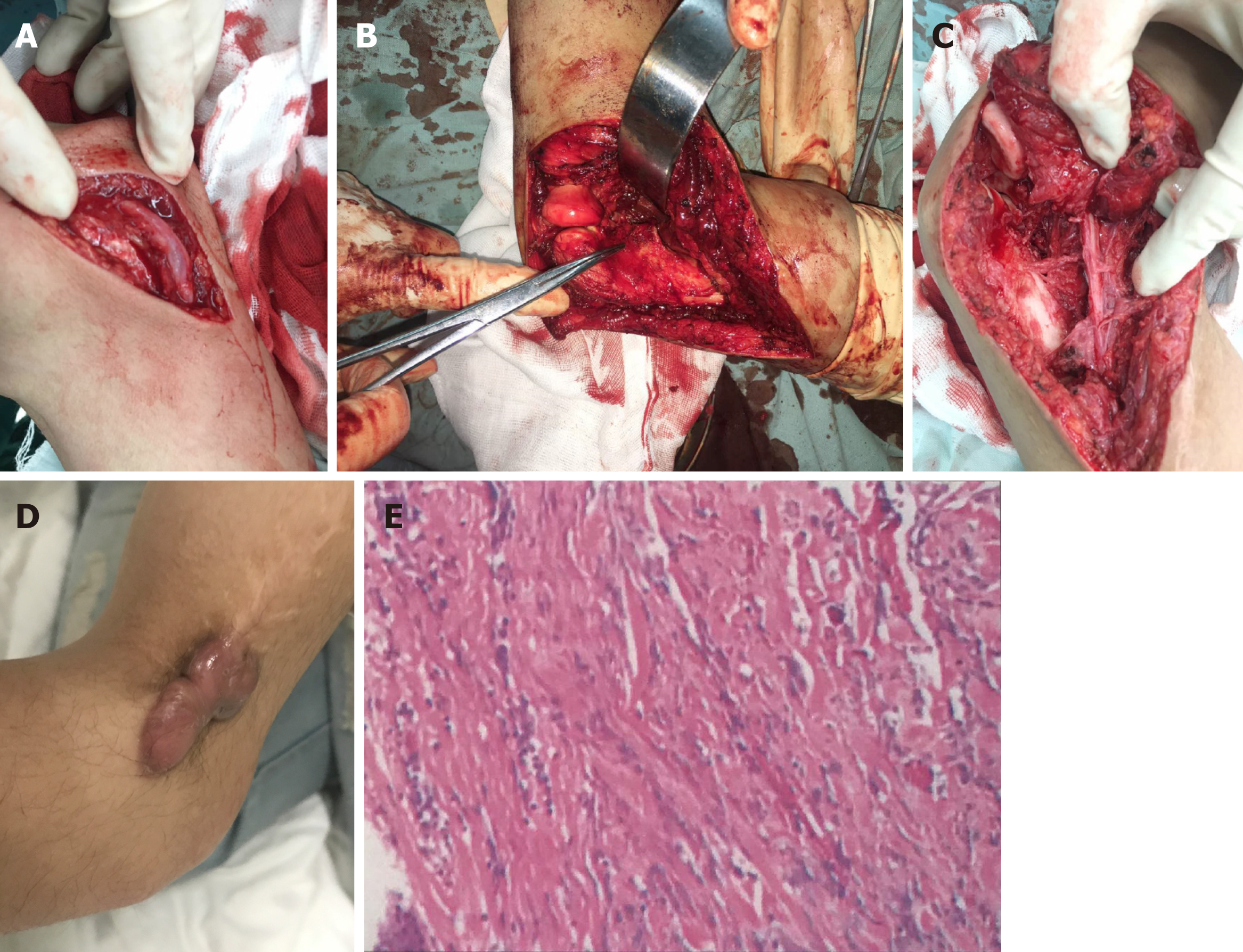
Figure 6 Detailed images of our surgery.
A: Intact ulnar nerve; B: Bony block of the humerus coronoid fossa; C: Radial nerve contracture; D: Scar of skin; E: Pathological section of the scar.
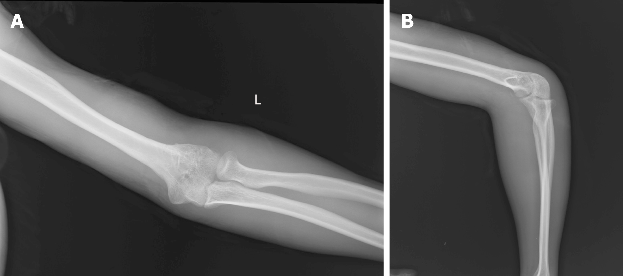
Figure 7 Radiography after the third operation.
A: Frontal x-ray of the left elbow; B: Lateral x-ray of the left elbow.
- Citation: Pan BQ, Huang J, Ni JD, Yan MM, Xia Q. Multiple rare causes of post-traumatic elbow stiffness in an adolescent patient: A case report and review of literature. World J Clin Cases 2019; 7(10): 1191-1199
- URL: https://www.wjgnet.com/2307-8960/full/v7/i10/1191.htm
- DOI: https://dx.doi.org/10.12998/wjcc.v7.i10.1191


