Copyright
©The Author(s) 2019.
World J Clin Cases. May 26, 2019; 7(10): 1177-1183
Published online May 26, 2019. doi: 10.12998/wjcc.v7.i10.1177
Published online May 26, 2019. doi: 10.12998/wjcc.v7.i10.1177
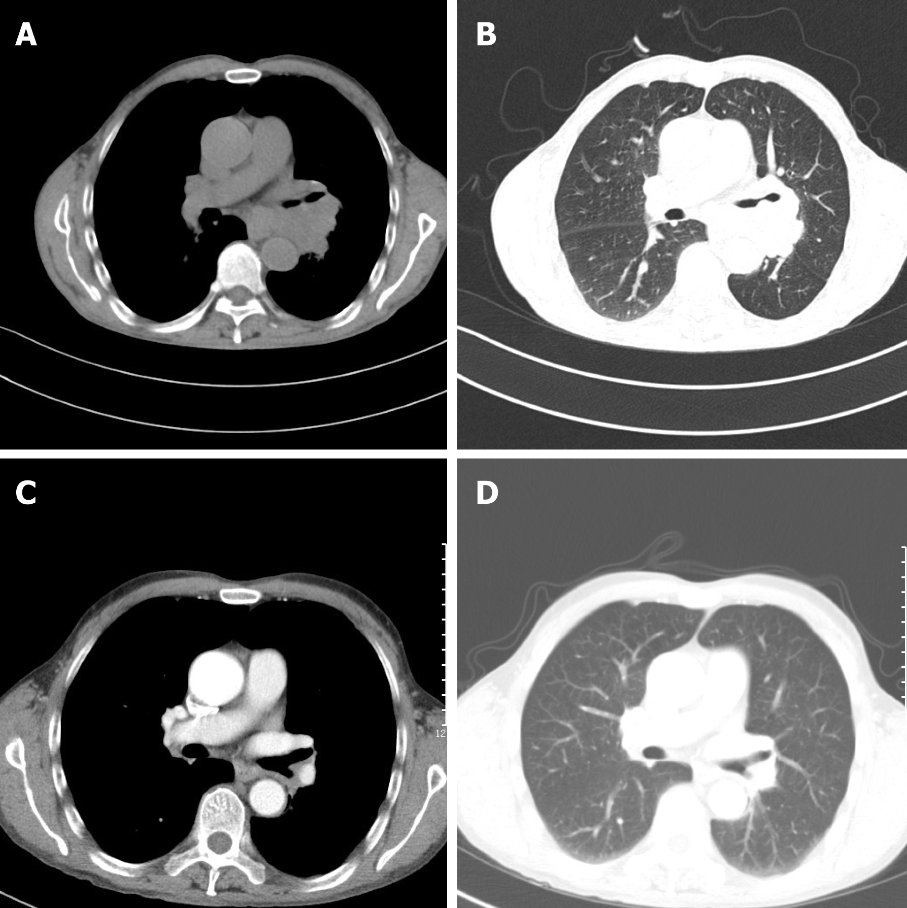
Figure 1 Computed tomography scan of the lung.
A, B: Chest computed tomography (CT) images obtained before chemotherapy indicated a central lung cancer was present. A lobulated, low density hilar mass (indicated with arrows) was found to be pressing and narrowing the left main bronchus. No lymph node enlargement was seen in the mediastinum; C, D: Chest CT images obtained after three rounds of chemotherapy. The left hilar mass (indicated with arrows) exhibited a marked reduction in size.
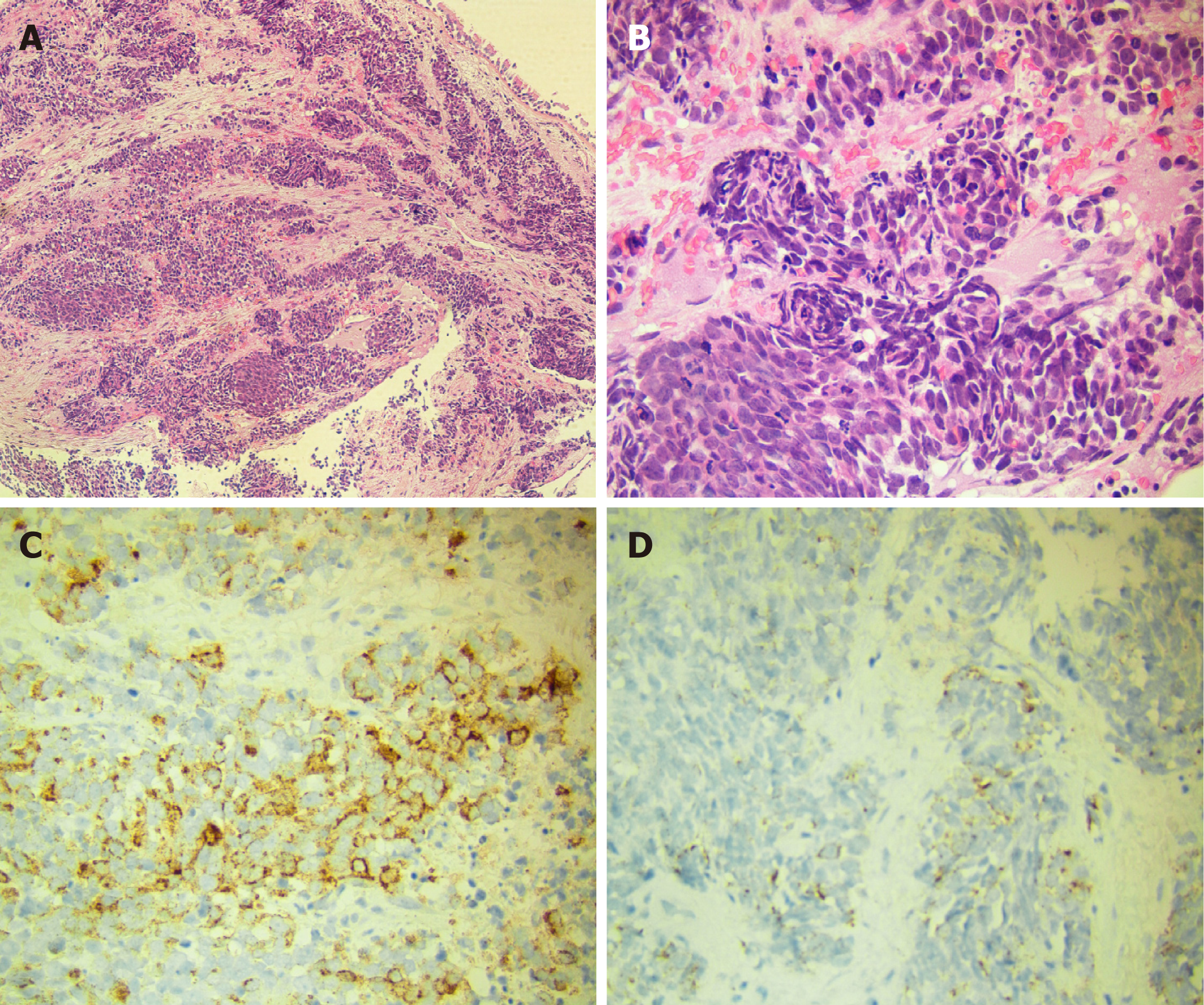
Figure 2 Immunohistochemical stainings of the hilar mass tissue biopsy sample.
A, B: HE staining detected small cells with hyperchromatic nuclei and minimal cytoplasm. Both spindly and lymphocytoid cancer cells were observed. Magnification, 100× and 400×, respectively; C, D: Immunohistochemical staining was also performed and focal positive staining for chromogranin A (C) and adrenocorticotropic hormone (D) were detected.
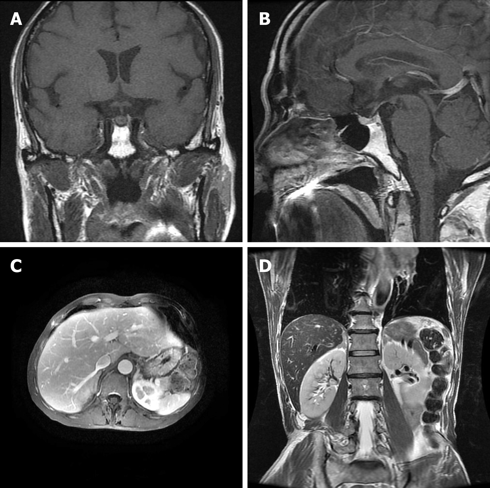
Figure 3 Magnetic resonance images of the head and the epigastrium.
A, B: Magnetic resonance imaging (MRI) of the pituitary. No obvious abnormalities were observed. Arrows are labeling the pituitary in both views; C, D: MRI of the epigastrium with bilateral adrenal thickening detected. Arrows are labeling the epigastrium in both views.
- Citation: Jin T, Wu F, Sun SY, Zheng FP, Zhou JQ, Zhu YP, Wang Z. Small cell lung cancer with panhypopituitarism due to ectopic adrenocorticotropic hormone syndrome: A case report. World J Clin Cases 2019; 7(10): 1177-1183
- URL: https://www.wjgnet.com/2307-8960/full/v7/i10/1177.htm
- DOI: https://dx.doi.org/10.12998/wjcc.v7.i10.1177


