Copyright
©The Author(s) 2019.
World J Clin Cases. May 26, 2019; 7(10): 1169-1176
Published online May 26, 2019. doi: 10.12998/wjcc.v7.i10.1169
Published online May 26, 2019. doi: 10.12998/wjcc.v7.i10.1169
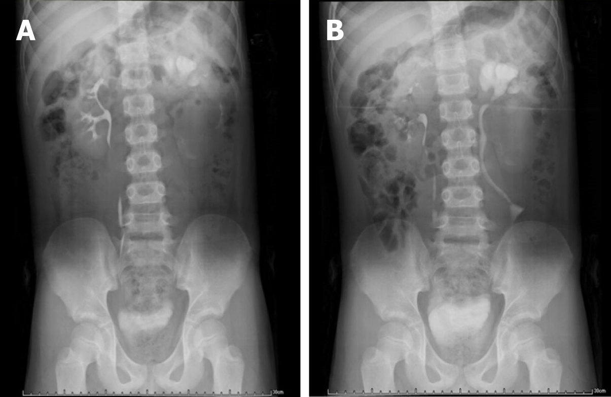
Figure 1 Intravenous pyelography.
A: 10 min, B: 40 min.
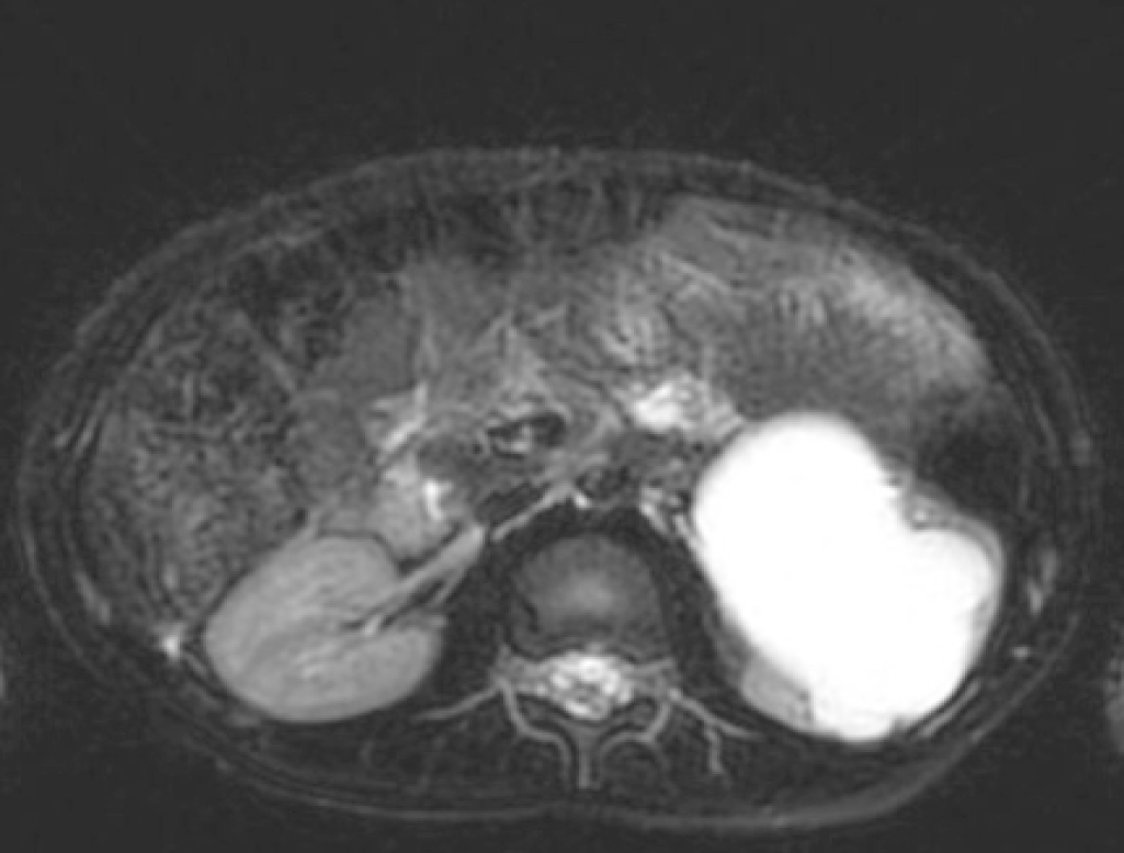
Figure 2 Magnetic resonance imaging of the patient (T2).
The left renal pelvis and ureter were significantly dilated, the widest part of the ureter was approximately 29 mm, and the left ureter had stenosis in the middle part.
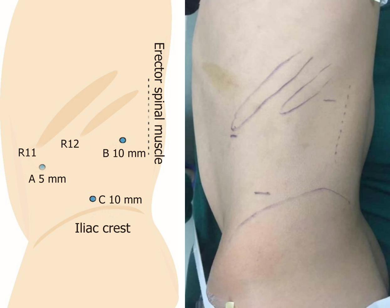
Figure 3 Trocar diagram.
Point C is the endoscopic port.
- Citation: Chen DX, Wang ZH, Wang SJ, Zhu YY, Li N, Wang XQ. Retroperitoneoscopic approach for partial nephrectomy in children with duplex kidney: A case report. World J Clin Cases 2019; 7(10): 1169-1176
- URL: https://www.wjgnet.com/2307-8960/full/v7/i10/1169.htm
- DOI: https://dx.doi.org/10.12998/wjcc.v7.i10.1169


