Copyright
©The Author(s) 2019.
World J Clin Cases. May 26, 2019; 7(10): 1149-1154
Published online May 26, 2019. doi: 10.12998/wjcc.v7.i10.1149
Published online May 26, 2019. doi: 10.12998/wjcc.v7.i10.1149
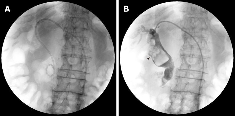
Figure 1 Cholangiography images in percutaneous transhepatic biliary drainage.
A: A 6 Fr pig-tail extra drainage tube is inserted; B: Black arrow head shows huge common bile duct stones (over 50 mm).
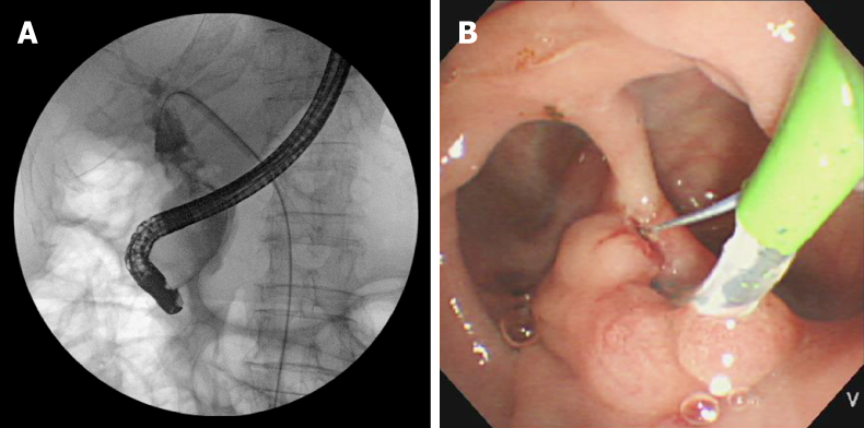
Figure 2 Images of endoscopic retrograde cholangiography and endoscopic sphincterotomy with rendezvous technique.
A: Cholangiography image of endoscopic retrograde cholangiography with rendezvous technique; B: Endoscopic image of endoscopic sphincterotomy. Large periampullary diverticula surrounding the papilla of Vater.
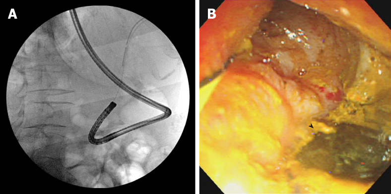
Figure 3 Images of direct peroral cholangioscopy by nasal endoscope with rendezvous technique.
A: Cholangiography images of insertion of nasal endoscope in common bile duct (CBD) by rendezvous technique; B: Direct peroral cholangioscopy image by nasal endoscope in CBD. Black arrow head shows adhered stone piece in the CBD wall.
- Citation: Kimura K, Kudo K, Yoshizumi T, Kurihara T, Yoshiya S, Mano Y, Takeishi K, Itoh S, Harada N, Ikegami T, Ikeda T. Electrohydraulic lithotripsy and rendezvous nasal endoscopic cholangiography for common bile duct stone: A case report. World J Clin Cases 2019; 7(10): 1149-1154
- URL: https://www.wjgnet.com/2307-8960/full/v7/i10/1149.htm
- DOI: https://dx.doi.org/10.12998/wjcc.v7.i10.1149


