Copyright
©The Author(s) 2019.
World J Clin Cases. May 26, 2019; 7(10): 1133-1141
Published online May 26, 2019. doi: 10.12998/wjcc.v7.i10.1133
Published online May 26, 2019. doi: 10.12998/wjcc.v7.i10.1133
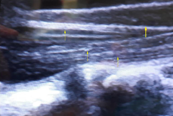
Figure 1 The conus medullaris and caud equina as seen on transdural ultrasound (US) examination.
The conus is hypoechogenic on US (thick arrow) and the cauda is hyperechogenic (thin arrows).
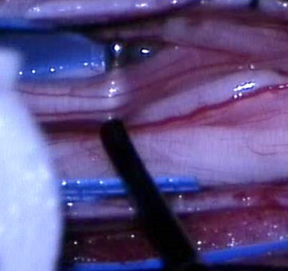
Figure 2 The exposure and isolation of the L1 root.
The root is composed of three fascicles, which will be subsequently separated. The motor fascicle will be spared, as will be one of the two sensory fascicles. The electrophysiological probe can be seen (right) and the cotton patty (left), which is used for isolation of the L1 root from other nerves.
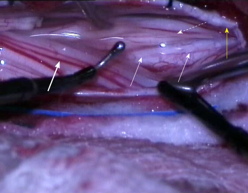
Figure 3 The intraoperative separation of motor and sensory roots and the identification of the groove that divides the roots anatomically.
The electrophysiological probe can be seen in place (right) as well as the groove on conus medullaris, separating the both groups of roots (thin arrows). The thick arrow indicates sensory roots and the dotted arrow indicates motor roots. The yellow arrow indicates the dural rim, held in place by tack-up sutures.
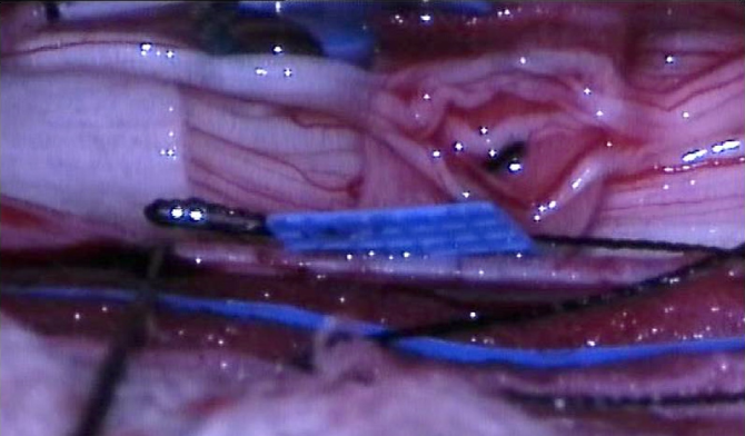
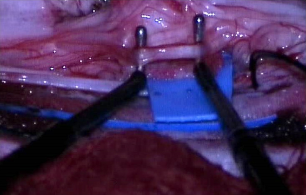
Figure 4 The separation of the sensory roots with an elastic cloth.
This root was again separated from others and divided into fascicles with a fine blunt microdisector.
Figure 5 The separation of the root into fascicles and electrophysiological monitoring for confirmation with both probes in place before the fascicle disconnection.
- Citation: Velnar T, Spazzapan P, Rodi Z, Kos N, Bosnjak R. Selective dorsal rhizotomy in cerebral palsy spasticity - a newly established operative technique in Slovenia: A case report and review of literature. World J Clin Cases 2019; 7(10): 1133-1141
- URL: https://www.wjgnet.com/2307-8960/full/v7/i10/1133.htm
- DOI: https://dx.doi.org/10.12998/wjcc.v7.i10.1133


