Copyright
©The Author(s) 2018.
World J Clin Cases. Nov 6, 2018; 6(13): 707-715
Published online Nov 6, 2018. doi: 10.12998/wjcc.v6.i13.707
Published online Nov 6, 2018. doi: 10.12998/wjcc.v6.i13.707
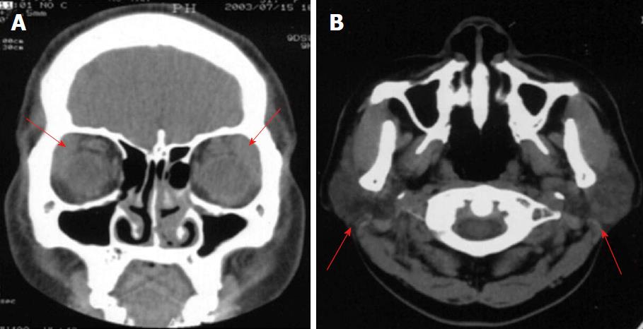
Figure 1 Involvement of the lacrimal gland parotid gland.
A: Computed tomography (CT) showing double lacrimal gland swelling on both sides; B: CT showing enlargement of the parotid gland.
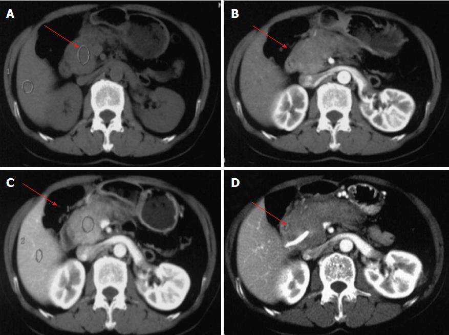
Figure 2 Involvement of pancreas and bile duct.
Abdominal computed tomography (CT) showed enlargement of the head of the pancreas. A: Plain scan showing the enlargement of the head of pancreas; B: Arterial phase showed a slight homogeneous enlargement; C: Portal phase showed an obvious enlargement of the pancreas; D: CT showed the bile duct stricture.
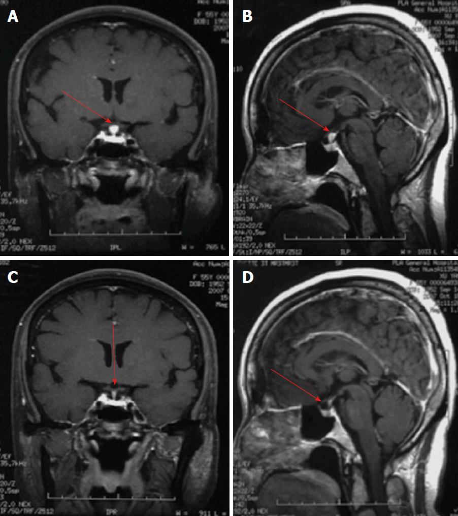
Figure 3 Involvement of the pituitary gland.
Magnetic resonance imaging showed the change of pituitary stalk nodular thickening. A: Coronal view clearly showed pituitary stalk nodular thickening (arrow); B: Sagittal view clearly showed pituitary stalk nodular thickening (arrow); C: Coronal view showing a significantly reduced pituitary stalk (arrow); D: Sagittal view showed a significantly reduced pituitary stalk (arrow).
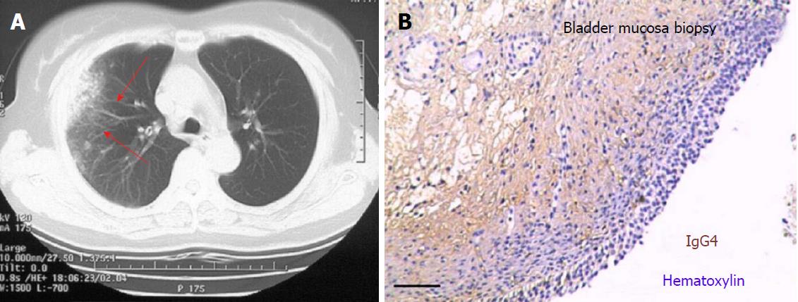
Figure 4 Involvement of lung and bladder mucosa.
A: Chest computed tomography showing interstitial pulmonary lesions (arrows) in the lung; B: Immunohistochemical (IHC) staining of IgG4 showed detectable IgG protein and IgG4-positive cell infiltration in bladder mocosa biopsy (bar = 50 μm).
- Citation: Xue J, Wang XM, Li Y, Zhu L, Liu XM, Chen J, Chi SH. Highlighting the importance of early diagnosis in progressive multi-organ involvement of IgG4-related disease: A case report and review of literature. World J Clin Cases 2018; 6(13): 707-715
- URL: https://www.wjgnet.com/2307-8960/full/v6/i13/707.htm
- DOI: https://dx.doi.org/10.12998/wjcc.v6.i13.707


