Copyright
©The Author(s) 2018.
World J Clin Cases. Nov 6, 2018; 6(13): 694-702
Published online Nov 6, 2018. doi: 10.12998/wjcc.v6.i13.694
Published online Nov 6, 2018. doi: 10.12998/wjcc.v6.i13.694
Figure 1 18F-fluorodeoxyglucose-positron emission tomography/computed tomography.
A: A swollen lymph node in the left submandibular region accompanied by increased fluorodeoxyglucose metabolism; B: Multiple lesions in the skeleton and the abdominal cavity with abnormally high metabolic activity; C: Swelling of the liver and spleen with increased metabolic activity accompanied by nodes with high metabolic activity in the parenchyma.
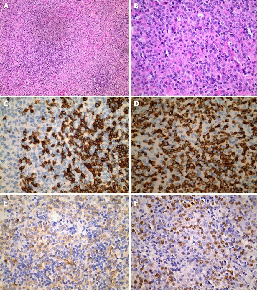
Figure 2 Histological findings.
A and B: Lymph node biopsy showing diffuse infiltration of malignant lymphoid cells (A: HE, ×100, B: ×400). Immunohistochemical staining for (C) CD20(+), (D) CD3(+), and (E) CD56(+) (× 400). F: In situ hybridization showing EBV-encoded small RNA positivity, with most cells showing strongly positive staining (×400). HE: Hematoxylin and eosin; EBV: Epstein-Barr virus.
- Citation: Liu QB, Zheng R. Natural killer/T-cell lymphoma with concomitant syndrome of inappropriate antidiuretic hormone secretion: A case report and review of literature. World J Clin Cases 2018; 6(13): 694-702
- URL: https://www.wjgnet.com/2307-8960/full/v6/i13/694.htm
- DOI: https://dx.doi.org/10.12998/wjcc.v6.i13.694


