Copyright
©The Author(s) 2018.
World J Clin Cases. Sep 26, 2018; 6(10): 398-405
Published online Sep 26, 2018. doi: 10.12998/wjcc.v6.i10.398
Published online Sep 26, 2018. doi: 10.12998/wjcc.v6.i10.398
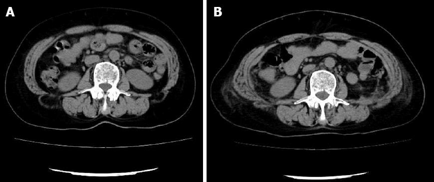
Figure 1 Preoperative vs postoperative imaging.
Comparison of the preoperative (A) and postoperative (B) images in a patient with a bilateral lumbar hernia.
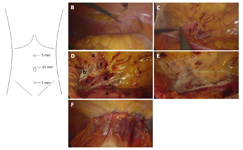
Figure 2 Surgical technique keynotes.
A: The location of trocars; B: The incision into the peritoneum; C: The exposure of hernia contents; D: The structure of the defect area. a: The twelfth rib; b: Neurovascular bundles; c: The erector spinae; d: The ilioinguinal nerve; e: The psoas major; f: The defect; E: The implantation of the meshes; F: An overview upon finishing the repair.
- Citation: Huang DY, Pan L, Chen MY, Fang J. Laparoscopic repair via the transabdominal preperitoneal procedure for bilateral lumbar hernia: Three cases report and review of literature. World J Clin Cases 2018; 6(10): 398-405
- URL: https://www.wjgnet.com/2307-8960/full/v6/i10/398.htm
- DOI: https://dx.doi.org/10.12998/wjcc.v6.i10.398


