Copyright
©The Author(s) 2017.
World J Clin Cases. Mar 16, 2017; 5(3): 119-123
Published online Mar 16, 2017. doi: 10.12998/wjcc.v5.i3.119
Published online Mar 16, 2017. doi: 10.12998/wjcc.v5.i3.119
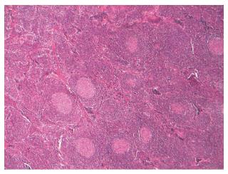
Figure 1 Light microscopy (HE × 400) demonstrates that reactive lymphoid follicles and the reactive germinal centers are radially penetrated by blood vessels in the lymph node specimen.
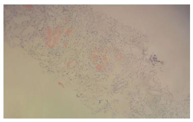
Figure 2 Light microscopy (HE × 400) demonstrates Congo red positive amyloid deposits in a glomerulus in the renal biopsy specimen.
- Citation: Eroglu E, Kocyigit I, Unal A, Sipahioglu MH, Akgun H, Kaynar L, Tokgoz B, Oymak O. Unicentric Castleman’s disease associated with end stage renal disease caused by amyloidosis. World J Clin Cases 2017; 5(3): 119-123
- URL: https://www.wjgnet.com/2307-8960/full/v5/i3/119.htm
- DOI: https://dx.doi.org/10.12998/wjcc.v5.i3.119


