Copyright
©The Author(s) 2017.
World Journal of Clinical Cases. Nov 16, 2017; 5(11): 390-396
Published online Nov 16, 2017. doi: 10.12998/wjcc.v5.i11.390
Published online Nov 16, 2017. doi: 10.12998/wjcc.v5.i11.390
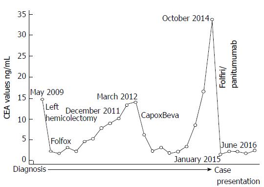
Figure 1 Time course of carcinoembryonic antigen levels during therapy and follow-up.
CEA: Carcinoembryonic antigen.
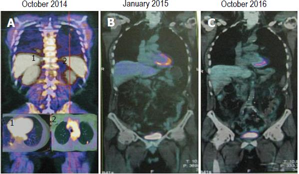
Figure 2 Positron emission tomography/computed tomography restaging after panitumumab-based therapy.
A: Positron emission tomography/computed tomography scan revealing two lung metastases; B: Complete response after 6 cycles of Folfiri/Panitumumab; C: Long-lasting complete response after maintenance therapy with panitumumab single agent.
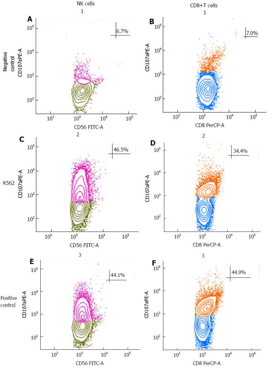
Figure 3 Cytotoxicity tests.
NK-cell mediated cytotoxicity was evaluated using the degranulation lysosomal marker CD107. The cytotoxic activity of NK cells was tested against NK-sensitive cell line K562. Medium alone served as the negative control and the positive control were NK cells stimulated with phorbol-12-myristate-13-acetate (PMA) and ionomycin, in presence of PE-conjugate anti-CD107a antibody. Control samples were incubated without target cells to detect spontaneous degranulation. NK cells were defined as CD3-CD56+ in the lymphocyte gate (A, C, E). CD8 cytotoxic T cells were analyzed in B, D and F panels (see Methods for details).
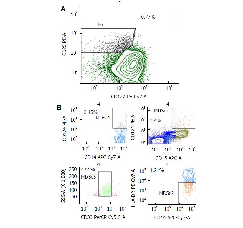
Figure 4 Characterization of regulatory cells.
Flow cytometry was performed on fresh venous blood; A: Identification of circulating T regulatory cells (Treg) on CD4+ cells (gate on low CD127 and high CD25). B: Identification of four types circulating myeloid-derived suppressor cells (MDSCs1, 2, 3, 4). Percentages are relative to peripheral blood lymphocytes.
- Citation: Ottaiano A, Napolitano M, Capozzi M, Tafuto S, Avallone A, Scala S. Natural killer cells activity in a metastatic colorectal cancer patient with complete and long lasting response to therapy. World Journal of Clinical Cases 2017; 5(11): 390-396
- URL: https://www.wjgnet.com/2307-8960/full/v5/i11/390.htm
- DOI: https://dx.doi.org/10.12998/wjcc.v5.i11.390


