Copyright
©The Author(s) 2016.
World J Clin Cases. Apr 16, 2016; 4(4): 118-123
Published online Apr 16, 2016. doi: 10.12998/wjcc.v4.i4.118
Published online Apr 16, 2016. doi: 10.12998/wjcc.v4.i4.118
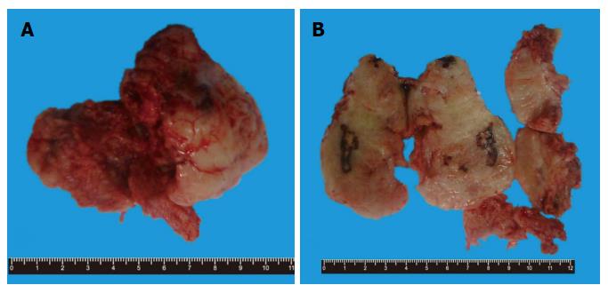
Figure 1 Gross findings of the specimen.
A: Elliptical gray-white and grey-red soft tissue mass with fibrous capsule; B: Medium texture with multiple hemorrhage and necrosis on the cut surface.
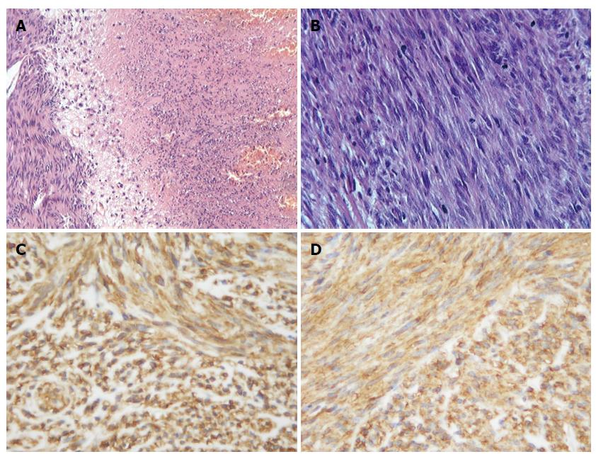
Figure 2 Histological findings of the tumor.
A: The tumor was composed of cellular spindle cells with a large area of necrosis. Hematoxylin and eosin (× 200); B: Active mitosis of tumor cells. Hematoxylin and eosin (× 400). Immunohistochemical findings of the tumors; C: Diffusely and strongly DOG1 positive in the tumor cells (× 400); D: Diffusely and strongly CD117 (c-kit pro-oncogene product) positive in the tumor cells (× 400).
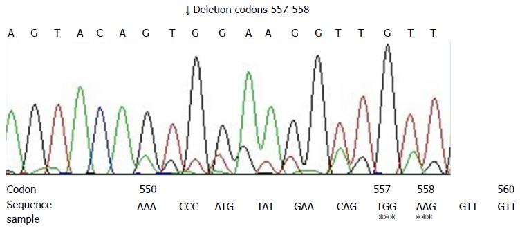
Figure 3 Computer analysis of a part of exon 11 of the c-kit gene in the tumor.
Deletion of codons 557-558 was noted. Asterisks showed the deletion.
- Citation: Liu QY, Kan YZ, Zhang MY, Sun TY, Kong LF. Primary extragastrointestinal stromal tumor arising in the vaginal wall: Significant clinicopathological characteristics of a rare aggressive soft tissue neoplasm. World J Clin Cases 2016; 4(4): 118-123
- URL: https://www.wjgnet.com/2307-8960/full/v4/i4/118.htm
- DOI: https://dx.doi.org/10.12998/wjcc.v4.i4.118


