Copyright
©The Author(s) 2015.
World J Clin Cases. Mar 16, 2015; 3(3): 327-329
Published online Mar 16, 2015. doi: 10.12998/wjcc.v3.i3.327
Published online Mar 16, 2015. doi: 10.12998/wjcc.v3.i3.327
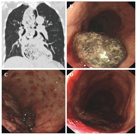
Figure 1 CT and endoscopy imaging.
A: CT showed lower esophageal dilatation and esophageal wall thickening; Upper endoscopy revealed; B: A large, dark round stone; C: Multiple ulcers on the esophageal wall; D: A slit in the cardiac mucosa with a large clot attached.
- Citation: Zhang WW, Xie XJ, Geng CX, Zhan SH. Rare case of upper gastrointestinal bleeding in achalasia. World J Clin Cases 2015; 3(3): 327-329
- URL: https://www.wjgnet.com/2307-8960/full/v3/i3/327.htm
- DOI: https://dx.doi.org/10.12998/wjcc.v3.i3.327


