Copyright
©The Author(s) 2015.
World J Clin Cases. Dec 16, 2015; 3(12): 988-992
Published online Dec 16, 2015. doi: 10.12998/wjcc.v3.i12.988
Published online Dec 16, 2015. doi: 10.12998/wjcc.v3.i12.988
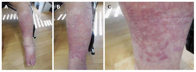
Figure 1 Distant (A), intermediate (B), and close (C) views of erythematous dermal plaques on the left leg of a 68-year-old man.
The lesions were later diagnosed as cutaneous sarcoid reaction.
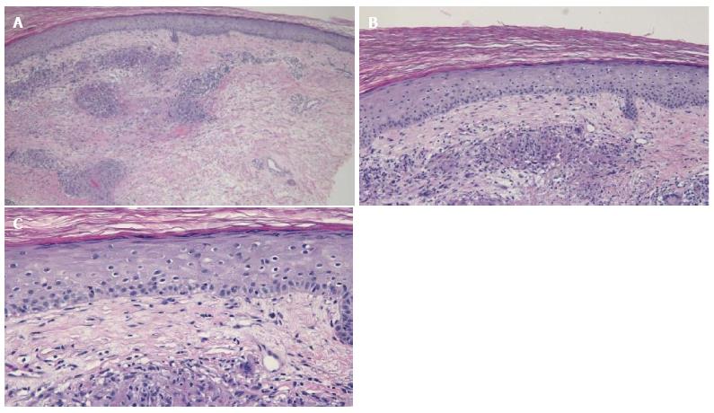
Figure 2 Low (A), intermediate (B), and high (C) magnification views of a sample of a lesion taken from the left leg of a 68-year-old man.
Multiple epithelioid granulomas can be observed in the superficial and mid-reticular dermis. Interstitial histiocytes can also be seen within the interstitium (hematoxylin and eosin: A: × 10; B: × 20; C: × 40).
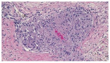
Figure 3 High magnification view of a sarcoidal granuloma from a sample of a lesion taken from the left leg of a 68-year-old man.
There is mild lymphocytic and neutrophilic inflammation surrounding the granuloma (hematoxylin and eosin: × 40).
- Citation: Beutler BD, Cohen PR. Sarcoma-associated sarcoid reaction: Report of cutaneous sarcoid reaction in a patient with liposarcoma. World J Clin Cases 2015; 3(12): 988-992
- URL: https://www.wjgnet.com/2307-8960/full/v3/i12/988.htm
- DOI: https://dx.doi.org/10.12998/wjcc.v3.i12.988


