Copyright
©The Author(s) 2015.
World J Clin Cases. Nov 16, 2015; 3(11): 942-945
Published online Nov 16, 2015. doi: 10.12998/wjcc.v3.i11.942
Published online Nov 16, 2015. doi: 10.12998/wjcc.v3.i11.942
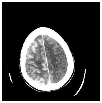
Figure 1 Non-contrast head computerized tomography showing extensive intravascular contrast with cortical staining, primarily over the right cerebral hemisphere.
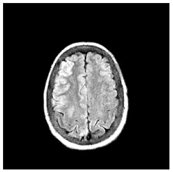
Figure 2 Magnetic resonance imaging fluid attenuated inversion recovery image showing hyperintense cortical signal of the cerebral hemispheres.
- Citation: Gollol Raju NS, Joshi D, Daggubati R, Movahed A. Contrast induced neurotoxicity following coronary angiogram with Iohexol in an end stage renal disease patient. World J Clin Cases 2015; 3(11): 942-945
- URL: https://www.wjgnet.com/2307-8960/full/v3/i11/942.htm
- DOI: https://dx.doi.org/10.12998/wjcc.v3.i11.942


