Copyright
©2014 Baishideng Publishing Group Inc.
World J Clin Cases. Sep 16, 2014; 2(9): 478-481
Published online Sep 16, 2014. doi: 10.12998/wjcc.v2.i9.478
Published online Sep 16, 2014. doi: 10.12998/wjcc.v2.i9.478
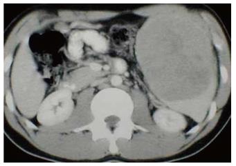
Figure 1 Computed tomography showing massive splenomegaly without mass lesion.
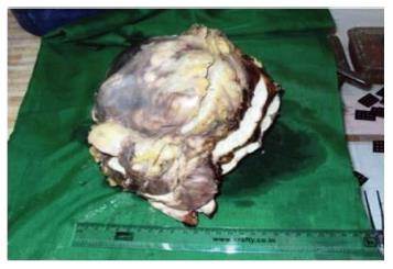
Figure 2 Encapsulated huge massive splenic mass.
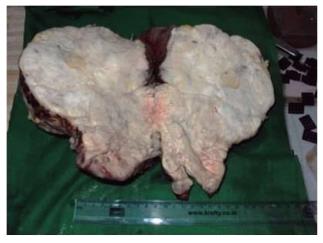
Figure 3 Cut surface was grey white.
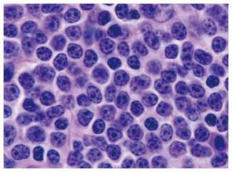
Figure 4 Diffuse monotonous population of neoplastic lymphoid cells.
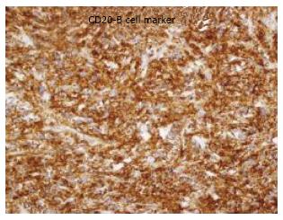
Figure 5 CD20 positive lymphoid cells.
- Citation: Ingle SB, Ingle CRH. Splenic lymphoma with massive splenomegaly: Case report with review of literature. World J Clin Cases 2014; 2(9): 478-481
- URL: https://www.wjgnet.com/2307-8960/full/v2/i9/478.htm
- DOI: https://dx.doi.org/10.12998/wjcc.v2.i9.478


