Copyright
©2014 Baishideng Publishing Group Inc.
World J Clin Cases. Sep 16, 2014; 2(9): 469-473
Published online Sep 16, 2014. doi: 10.12998/wjcc.v2.i9.469
Published online Sep 16, 2014. doi: 10.12998/wjcc.v2.i9.469
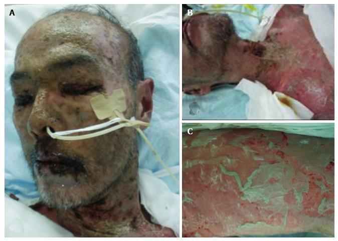
Figure 1 Skin findings in this patient.
A and B: Wide-spread blistering exanthemas of macules around the face and neck; C: Nikolsky phenomenon (slight rubbing of the skin resulting in exfoliation of the outermost layer) of the lower leg.
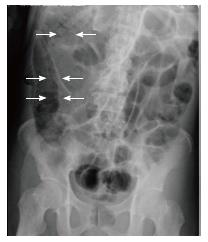
Figure 2 Plain supine abdominal radiography of patient on day 8 of admission to the intensive care unit, showing small-bowel distension and pneumatosis cystoides intestinalis (arrows).
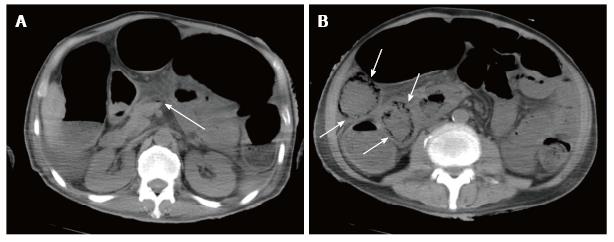
Figure 3 Abdominal computed tomography of patient on day 8 of admission to the intensive care unit, showing.
A: Gas in the superior mesenteric vein (arrow); B: Extraluminal gas along the small bowel mesentery (arrows).
- Citation: Yao SY, Seo R, Nagano T, Yamazaki K. Pneumatosis cystoides intestinalis associated with toxic epidermal necrolysis: A case report. World J Clin Cases 2014; 2(9): 469-473
- URL: https://www.wjgnet.com/2307-8960/full/v2/i9/469.htm
- DOI: https://dx.doi.org/10.12998/wjcc.v2.i9.469


