Copyright
©2014 Baishideng Publishing Group Inc.
World J Clin Cases. Jul 16, 2014; 2(7): 311-315
Published online Jul 16, 2014. doi: 10.12998/wjcc.v2.i7.311
Published online Jul 16, 2014. doi: 10.12998/wjcc.v2.i7.311
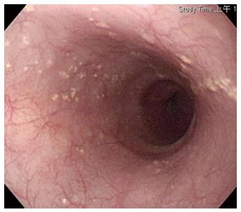
Figure 1 Sebaceous gland metaplasia in the esophagus.
Numerous tiny round yellowish lesions clustering distribution at the submucosa of the middle and lower esophagus.
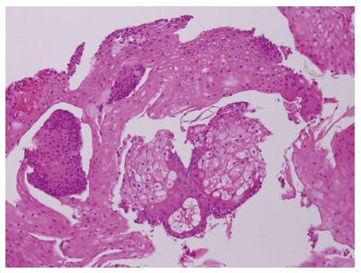
Figure 2 Esophageal squamous epithelial with sebaceous glands (HE stained × 400).
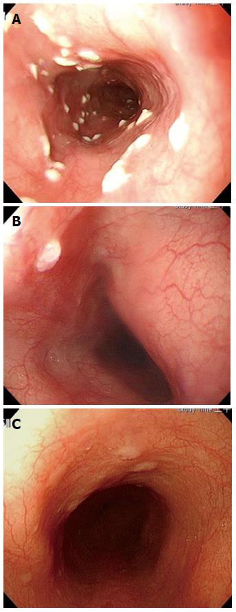
Figure 3 Photograph.
A: Candida infection of the esophagus. Multiple small brightly whitish elevated patches at the upper and middle esophagus; B: Papilloma of the esophagus. A single round whitish elevated nodule at the middle esophagus; C: Glycogenic acanthosis of the esophagus. Some small round lucent to lightly whitish nodules at the upper and middle esophagus.
- Citation: Chiu KW, Wu CK, Lu LS, Eng HL, Chiou SS. Diagnostic pitfall of sebaceous gland metaplasia of the esophagus. World J Clin Cases 2014; 2(7): 311-315
- URL: https://www.wjgnet.com/2307-8960/full/v2/i7/311.htm
- DOI: https://dx.doi.org/10.12998/wjcc.v2.i7.311


