Copyright
©2014 Baishideng Publishing Group Co.
World J Clin Cases. Feb 16, 2014; 2(2): 27-31
Published online Feb 16, 2014. doi: 10.12998/wjcc.v2.i2.27
Published online Feb 16, 2014. doi: 10.12998/wjcc.v2.i2.27
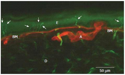
Figure 1 Epidermal nerve fibers (arrows) identified by immunofluorescence in the distal leg of a normal subject (in green, PGP 9.
5 staining of nerve fibers; in red, type IV collagen staining of basement membrane and blood vessels). E: Epidermis; D: Dermis; BM: Basement Membrane; A: Artery.
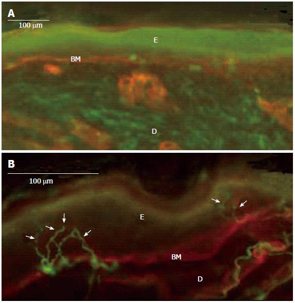
Figure 2 Complete denervation of epidermis (and dermis) in an 82-year-old patient with a severe post-herpetic neuralgia (A), normal, contralateral, mirror skin innervation (13 fibers/mm) (B).
Immunofluorescence method: In green, PGP 9.5 staining of nerve fibers; in red, type IV collagen staining of basement membrane and blood vessels; Arrows: Epidermal nerve fibers; E: Epidermis; D: Dermis; BM: Basement Membrane.
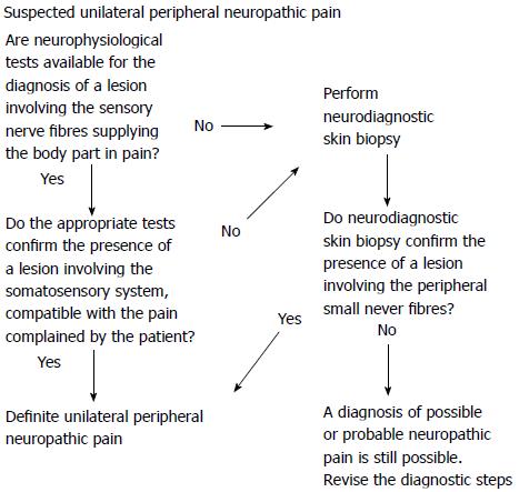
Figure 3 Suggested diagnostic sequences for the confirmation of a peripheral nerve lesion sustaining a definite, peripheral, unilateral neuropathic pain.
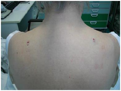
Figure 4 Figure exemplifies the bilateral biopsy needed for comparing the epidermal nerve fiber density of two symmetrical, mirror skin parts.
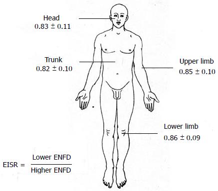
Figure 5 Figure shows the values of epidermal innervation symmetry ratio obtained for different body parts in 133 normal subjects.
ENFD: Epidermal Nerve Fiber Density.
- Citation: Buonocore M. Unilateral peripheral neuropathic pain: The role of neurodiagnostic skin biopsy. World J Clin Cases 2014; 2(2): 27-31
- URL: https://www.wjgnet.com/2307-8960/full/v2/i2/27.htm
- DOI: https://dx.doi.org/10.12998/wjcc.v2.i2.27


