Copyright
©2014 Baishideng Publishing Group Inc.
World J Clin Cases. Dec 16, 2014; 2(12): 888-892
Published online Dec 16, 2014. doi: 10.12998/wjcc.v2.i12.888
Published online Dec 16, 2014. doi: 10.12998/wjcc.v2.i12.888
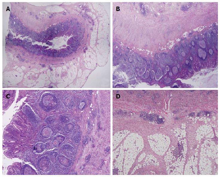
Figure 1 Appendiceal Crohn’s disease.
A: The appendix with Crohn’s disease shows transmural inflammation with markedly thickened wall; B: There is a prominent lymphoid hyperplasia in the mucosa and serosa; C: The mucosa shows many small non-caseating granulomas; D: The serosa shows creeping fat with perpendicular thick fibrous bands.
- Citation: Han H, Kim H, Rehman A, Jang SM, Paik SS. Appendiceal Crohn’s disease clinically presenting as acute appendicitis. World J Clin Cases 2014; 2(12): 888-892
- URL: https://www.wjgnet.com/2307-8960/full/v2/i12/888.htm
- DOI: https://dx.doi.org/10.12998/wjcc.v2.i12.888


