Copyright
©2014 Baishideng Publishing Group Inc.
World J Clin Cases. Nov 16, 2014; 2(11): 711-716
Published online Nov 16, 2014. doi: 10.12998/wjcc.v2.i11.711
Published online Nov 16, 2014. doi: 10.12998/wjcc.v2.i11.711
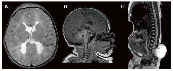
Figure 1 Pre-operative magnetic resonance imaging imaging of the patient to evaluate and screen for anatomical abnormalities.
A: Axial T2 MRI of the head at birth demonstrating no signs of hydrocephalus; B: Sagittal T1 MRI of the head and neck at birth with evidence of a tight cervicomedullary junction, a small posterior fossa, and hypoplastic corpus callosum and cerebellar vermis; C: Sagittal T2 MRI of the spine at birth demonstrating a large 7 cm × 7 cm sacral myelomeningocele with evidence of tethering. MRI: Magnetic resonance imaging.
Figure 2 T-2 weighted magnetic resonance imaging demonstrating shunt failure and progression of hydrocephalous over the course of 4 mo of treatment.
A: Pre-shunting axial T-2 weighted MRI ordered after patient had increased head circumference and suture splaying demonstrating ventriculomegaly; B, C: Subtle increase in ventricular size at 2 wk (B) and 6 wk post-shunting (C); D: At 4 mo post-operative shunting with significant increase in ventriculomegaly and extra axial fluid spacing. MRI: Magnetic resonance imaging.
- Citation: Awad AW, Aleck KA, Bhardwaj RD. Concomitant achondroplasia and Chiari II malformation: A double-hit at the cervicomedullary junction. World J Clin Cases 2014; 2(11): 711-716
- URL: https://www.wjgnet.com/2307-8960/full/v2/i11/711.htm
- DOI: https://dx.doi.org/10.12998/wjcc.v2.i11.711


