Copyright
©2014 Baishideng Publishing Group Co.
World J Clin Cases. Jan 16, 2014; 2(1): 1-4
Published online Jan 16, 2014. doi: 10.12998/wjcc.v2.i1.1
Published online Jan 16, 2014. doi: 10.12998/wjcc.v2.i1.1
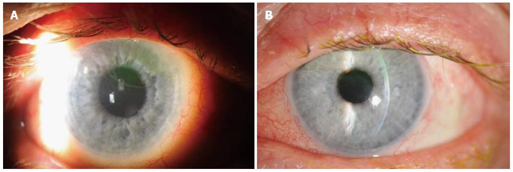
Figure 1 Photograph of right eye demonstrating epithelial demarcation line, fluorescein pooling, and ExPRESS shunt immediately below superior eyelid margin (A) and photograph of left eye showing epithelial demarcation line, superior stromal thinning, fluorescein pooling, and Ahmed valve immediately below superior eyelid margin (B).
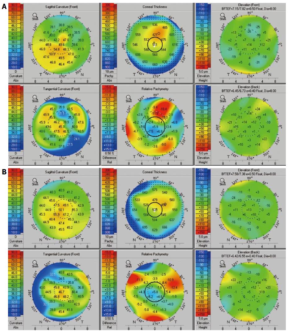
Figure 2 Oculus Pentacam of right eye (A), left eye (B) showing corneal thinning and irregular curvature.
- Citation: Fenzl CR, Moshirfar M, Gess AJ, Muthappan V, Goldsmith J. Dellen-like keratopathy associated with glaucoma drainage devices. World J Clin Cases 2014; 2(1): 1-4
- URL: https://www.wjgnet.com/2307-8960/full/v2/i1/1.htm
- DOI: https://dx.doi.org/10.12998/wjcc.v2.i1.1


