Copyright
©The Author(s) 2025.
World J Clin Cases. Mar 16, 2025; 13(8): 100028
Published online Mar 16, 2025. doi: 10.12998/wjcc.v13.i8.100028
Published online Mar 16, 2025. doi: 10.12998/wjcc.v13.i8.100028
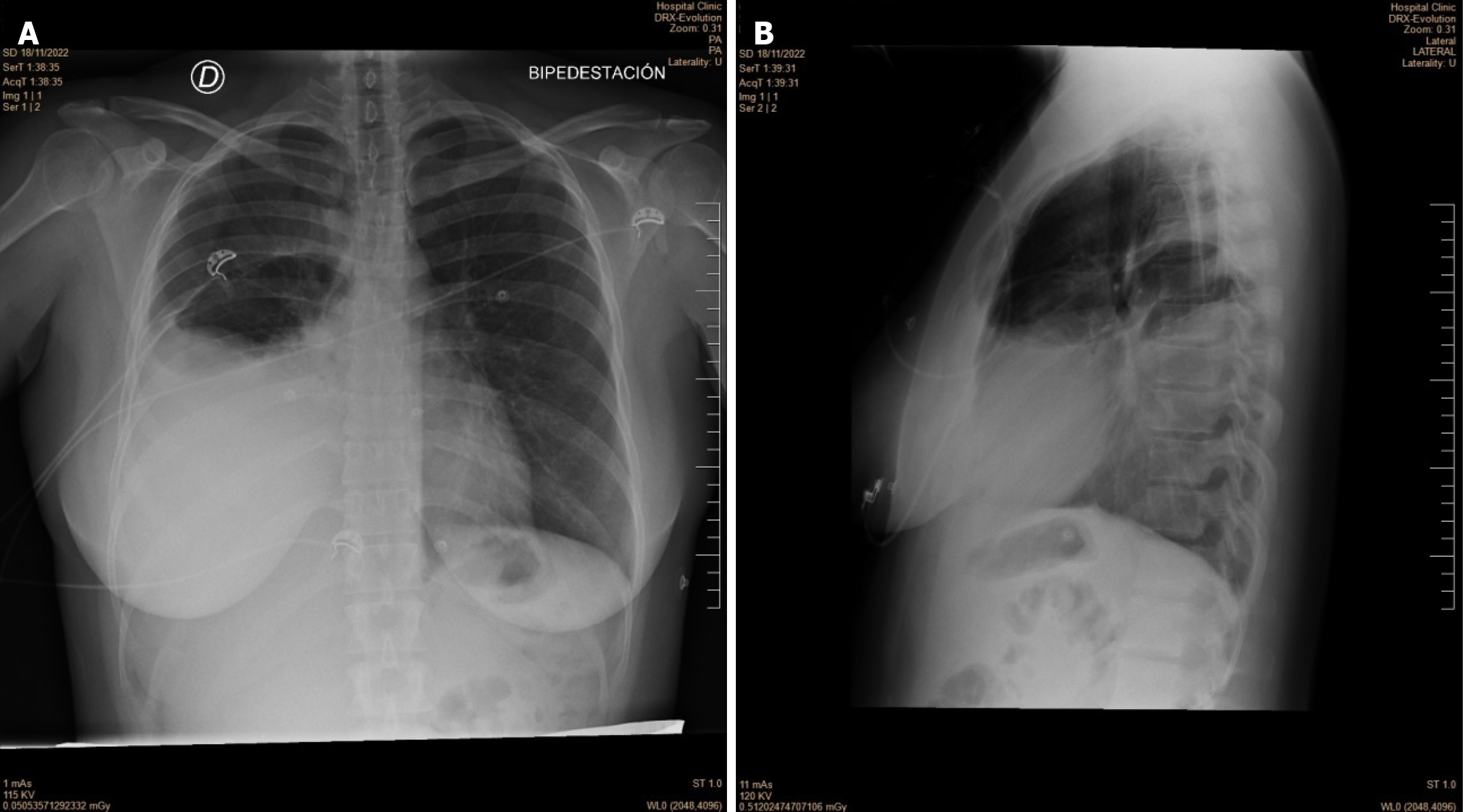
Figure 1 Chest X-ray upon admission (November 18, 2022) showing right pleural effusion.
A: Erect anteroposterior view; B. Erect lateral view.
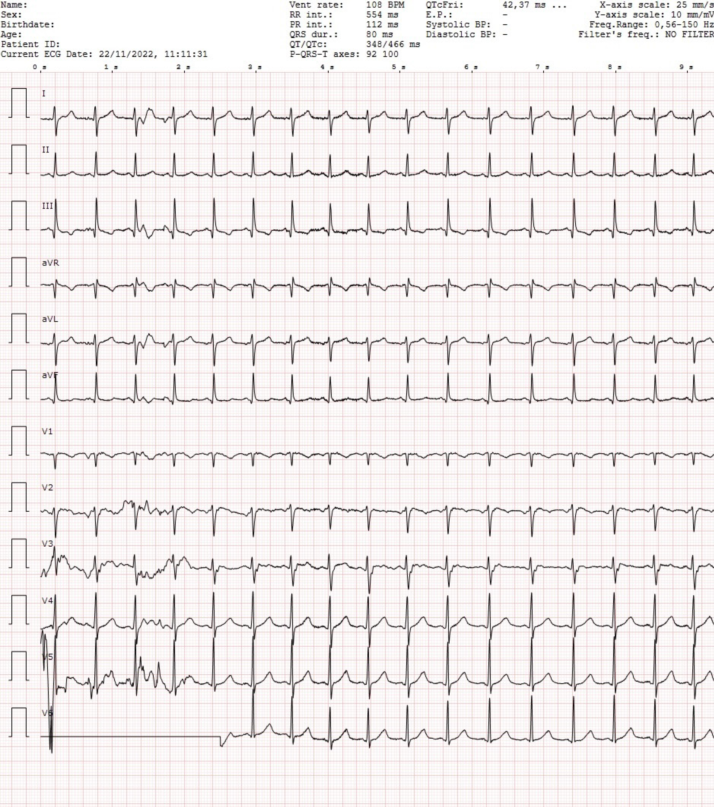
Figure 2
Screening electrocardiogram (November 22, 2022) showed sinus tachycardia, a slight right axis deviation of approximately 90°, and negative T in V1 and inferior leads.
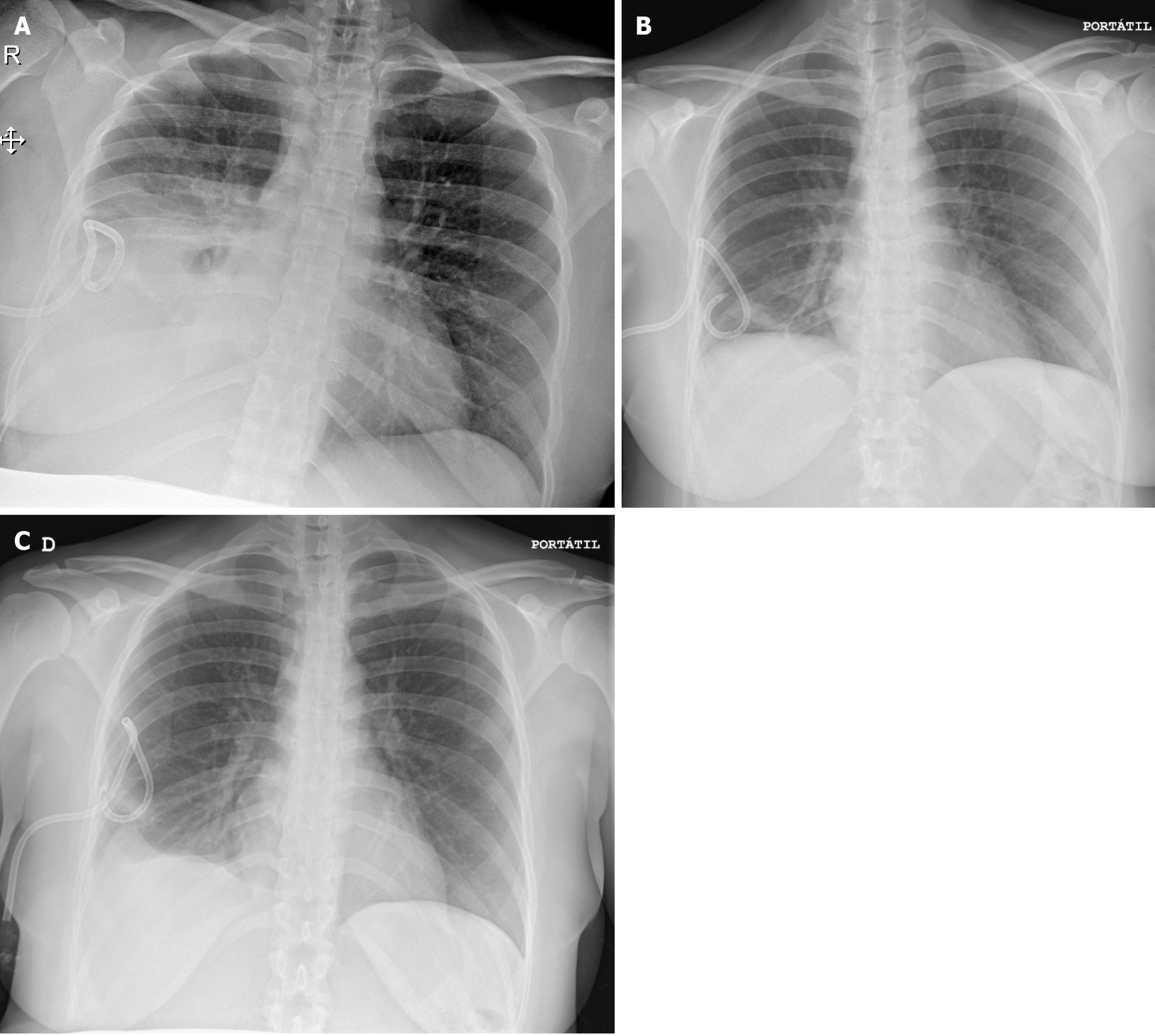
Figure 3 Chest X-ray.
A: Chest X-ray after ultrasound-guided thoracentesis (yielding a volume of 1200 mL) and placement of the pigtail catheter for continuous pleural drainage of the right pleural effusion (November 22, 2022); B: Chest X-ray follow-up (November 23, 2022). The evolution of the patient was favorable, with periodic clamping of the drainage and treatment with furosemide and albumin; C: Chest X-ray follow-up (November 26, 2022). On this day, treatment with furosemide and albumin was discontinued. The pleural drainage was removed two days after, having drained up to 5 L during the whole hospitalization period.
- Citation: Solsona Í, Peralta S, Barral Y, Fàbregues F, Giménez-Bonafé P. Unusual ovarian hyperstimulation syndrome presentation: Pleural effusion without ascites. A case report. World J Clin Cases 2025; 13(8): 100028
- URL: https://www.wjgnet.com/2307-8960/full/v13/i8/100028.htm
- DOI: https://dx.doi.org/10.12998/wjcc.v13.i8.100028


