Copyright
©The Author(s) 2025.
World J Clin Cases. Jul 26, 2025; 13(21): 105816
Published online Jul 26, 2025. doi: 10.12998/wjcc.v13.i21.105816
Published online Jul 26, 2025. doi: 10.12998/wjcc.v13.i21.105816
Figure 1 The electrocardiography shows anterior myocardial infarction.
A: The electrocardiography (ECG) at the time of admission, with a typical ST-segment elevation in leads V1–V5, indicating anterior myocardial infarction; B: Three hours after admission, a repeat ECG shows anterior ST-elevation myocardial infarction, with no significant difference from the initial ECG, suggesting the unsuccessful recanalization of the culprit coronary artery.
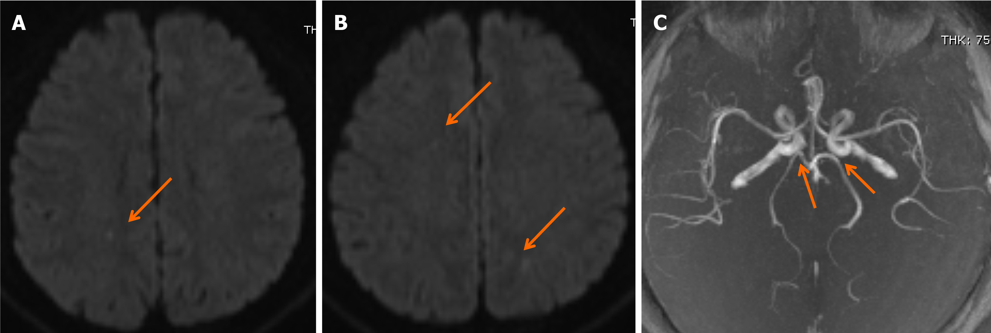
Figure 2 The magnetic resonance imaging shows acute ischemic stroke.
A and B: Diffusion-weighted magnetic resonance imaging reveals subtle scattered ischemic changes at the right frontal lobe and both parietal lobes; C: Magnetic resonance angiography shows an incomplete basilar artery ring and the absence of bilateral posterior communicating arteries.
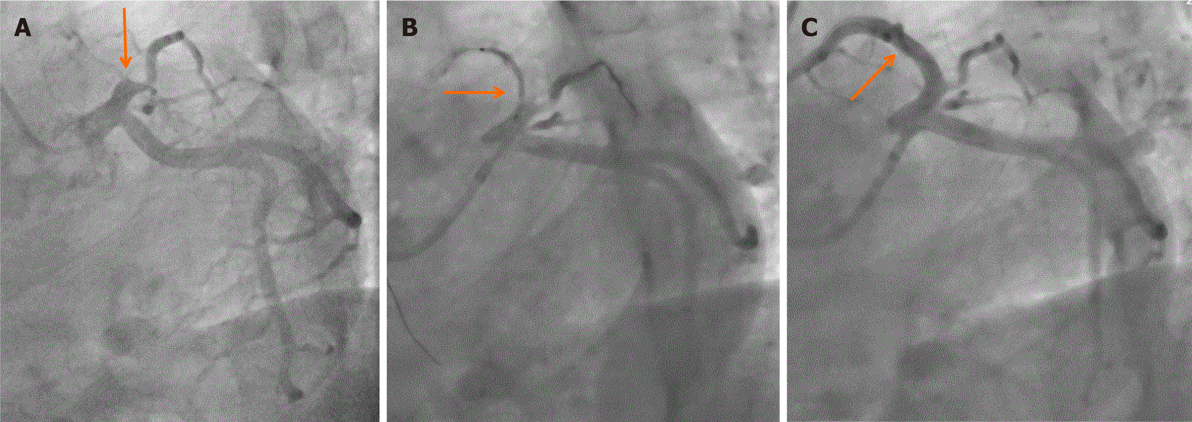
Figure 3 Percutaneous coronary intervention was successfully performed.
A: The coronary angiogram revealed 100% occlusion of the mid-left anterior descending (LAD) artery; B: Percutaneous transluminal coronary angioplasty was performed at the LAD artery with unsuccessful recanalization. The residual diameter stenosis was 80%-90%; C: After stenting, the coronary angiogram shows LAD artery with thrombolysis in myocardial infarction grade 3 flow.
- Citation: Zheng WX, Liu LY. Urgent thrombolysis followed by percutaneous coronary intervention for the simultaneous acute cardio-cerebral ischemic attack: A case report. World J Clin Cases 2025; 13(21): 105816
- URL: https://www.wjgnet.com/2307-8960/full/v13/i21/105816.htm
- DOI: https://dx.doi.org/10.12998/wjcc.v13.i21.105816


