Copyright
©The Author(s) 2025.
World J Clin Cases. Jul 26, 2025; 13(21): 103841
Published online Jul 26, 2025. doi: 10.12998/wjcc.v13.i21.103841
Published online Jul 26, 2025. doi: 10.12998/wjcc.v13.i21.103841
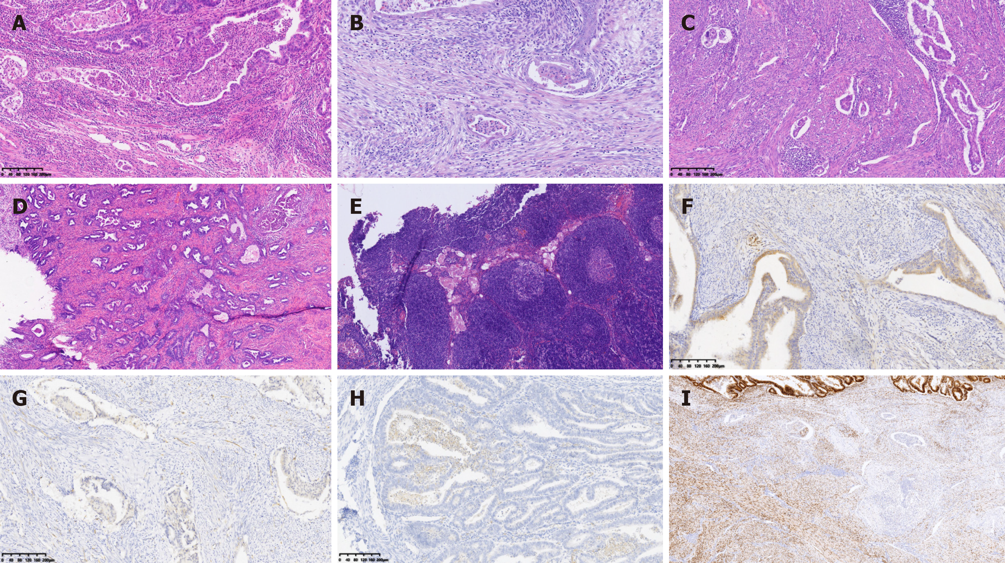
Figure 1 Integrated analysis of histopathological findings and Immunohistochemistry results.
A-C: Representative image of endometrioid endometrial carcinoma (EEC) with microcystic, elongated, and fragmented (MELF) (× 200); D: Representative image of EEC with MELF and cervical stromal involvement (× 40); E: Lymph node metastasis (× 200); F: Image showing strong positive CXC chemokine receptor 4 (CXCR4) expression in MELF area (× 200); G: Image showing weak positive CXCR4 expression in MELF area (× 200); H: Image showing weak positive CXCR4 expression in EEC main area (× 200); I: Image showing positive estrogen receptor expression in EEC, strong positive in the main area, weakly positive in MELF area (in the lower picture) (× 100).
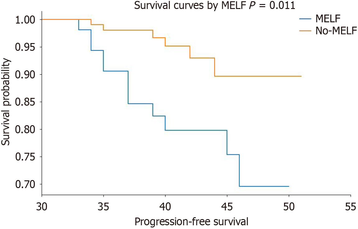
Figure 2 Survival estimate between microcystic, elongated, and fragmented group and non- microcystic, elongated, and fragmented group.
There was significant difference in progression-free survival between microcystic, elongated, and fragmented (MELF) group and non-MELF group by Kaplan-Meier analysis (P < 0.05). MELF: Microcystic, elongated, and fragmented.
- Citation: Wang JX, Shen DH, Zhang XB. CXCR4 expression and clinicopathological features of endometrial endometrioid carcinoma with the microcystic, elongated, and fragmented pattern. World J Clin Cases 2025; 13(21): 103841
- URL: https://www.wjgnet.com/2307-8960/full/v13/i21/103841.htm
- DOI: https://dx.doi.org/10.12998/wjcc.v13.i21.103841


