Copyright
©The Author(s) 2025.
World J Clin Cases. May 16, 2025; 13(14): 102791
Published online May 16, 2025. doi: 10.12998/wjcc.v13.i14.102791
Published online May 16, 2025. doi: 10.12998/wjcc.v13.i14.102791
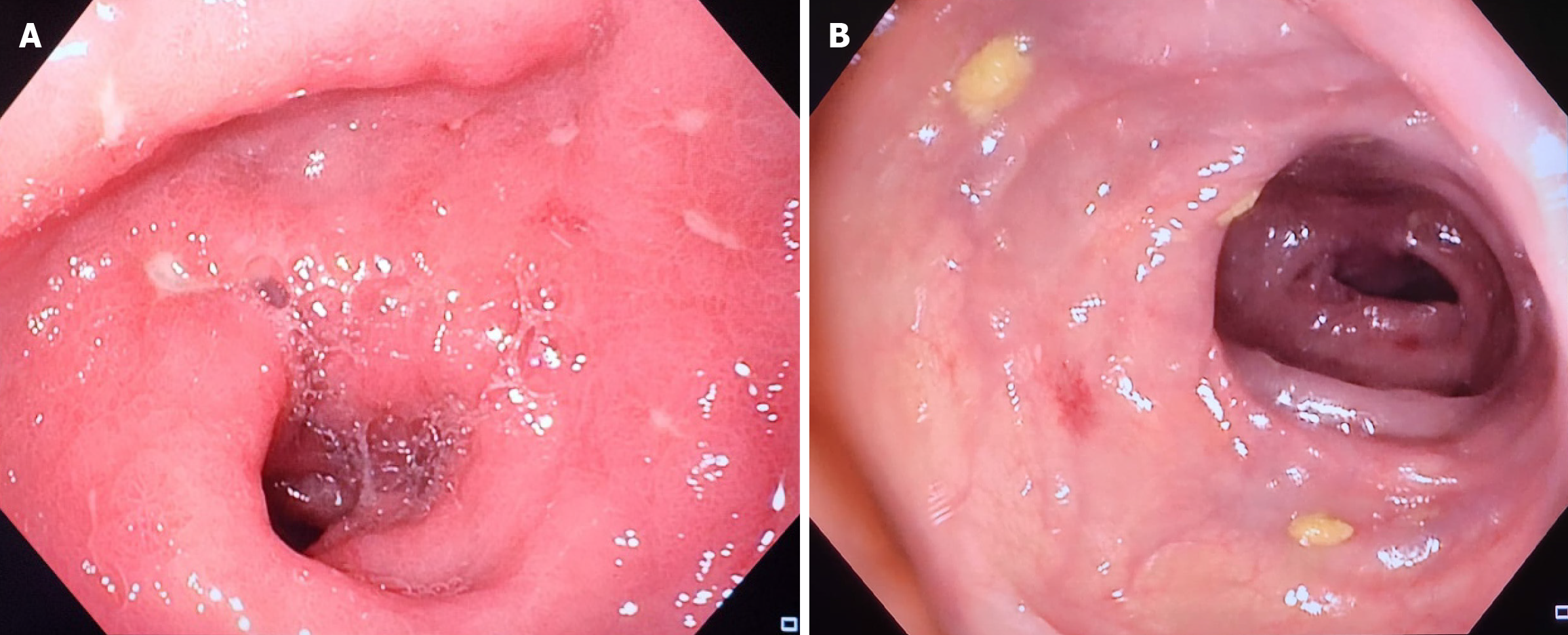
Figure 1 A colonoscopy view revealing multiple ulcerations at the level of the rectosigmoid area with surrounding erythematous and edematous mucosa.
A: Multiple superficial aphtous ulcerations noted at the level of the rectosigmoid junction; B: Erosive erythematous mucosa seen at the rectosigmoid junction.
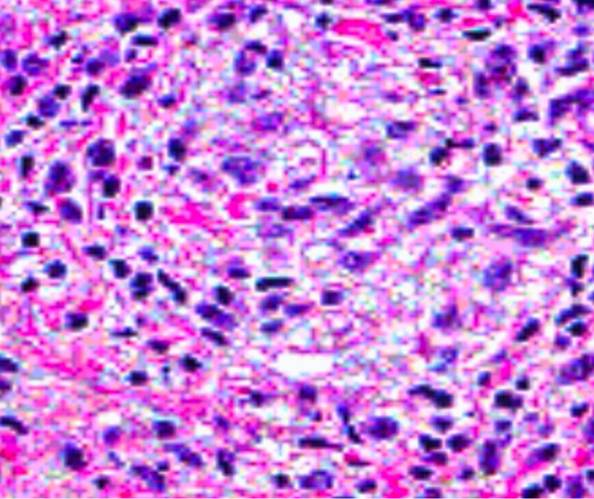
Figure 2
Hematoxylin and eosin stain revealing dense nodular inflammatory infiltrate comprised of plasma cells, neutrophils, lympho
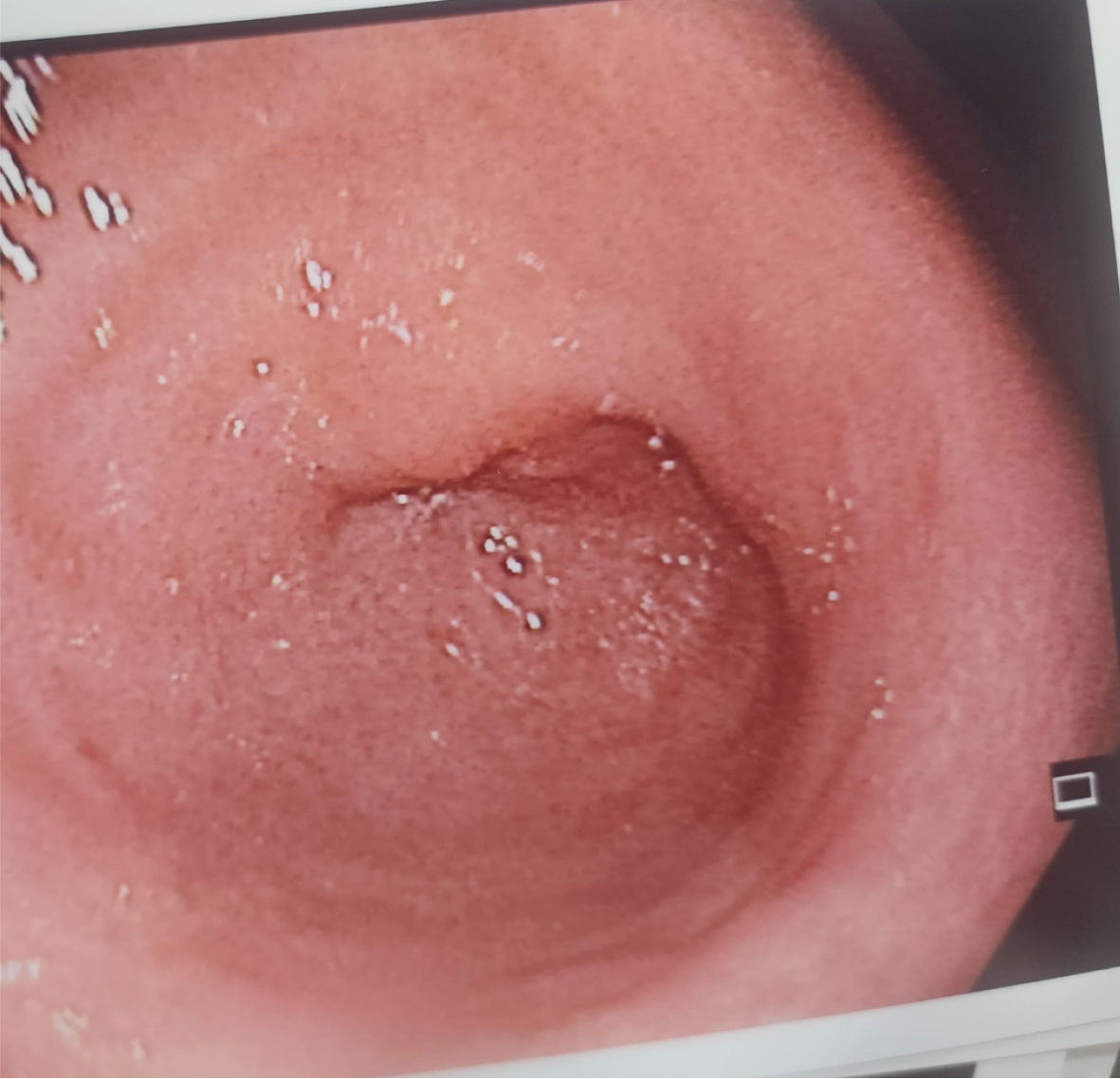
Figure 3
A repeat colonoscopy revealing normal rectosigmoid junction with normal healed mucosa.
- Citation: Khoury KM, Jradi A, Karam K, Fiani E. Lymphogranuloma venereum proctosigmoiditis misdiagnosed as inflammatory bowel disease: A case report. World J Clin Cases 2025; 13(14): 102791
- URL: https://www.wjgnet.com/2307-8960/full/v13/i14/102791.htm
- DOI: https://dx.doi.org/10.12998/wjcc.v13.i14.102791


