Copyright
©The Author(s) 2025.
World J Clin Cases. May 6, 2025; 13(13): 98937
Published online May 6, 2025. doi: 10.12998/wjcc.v13.i13.98937
Published online May 6, 2025. doi: 10.12998/wjcc.v13.i13.98937
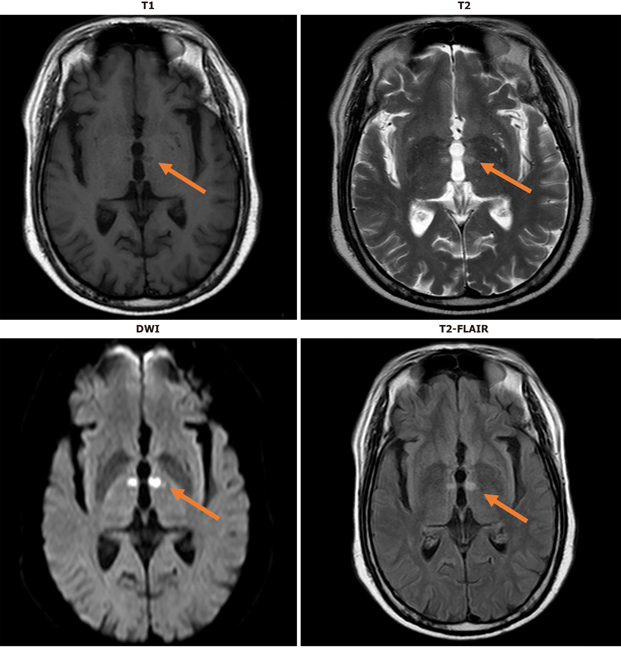
Figure 1 Magnetic resonance imaging.
This figure clearly reflects the imaging manifestations of acute cerebral infarction closely associated with Percheron artery occlusion on various magnetic resonance imaging sequences, including but not limited to T1-weighted imaging, T2-weighted imaging, diffusion-weighted imaging, and fluid-attenuated inversion-recovery sequences. Each sequence provides unique information regarding the location, extent, and characteristics of the infarcted area within the brain. DWI: Diffusion-weighted imaging; FLAIR: Fluid-attenuated inversion-recovery.
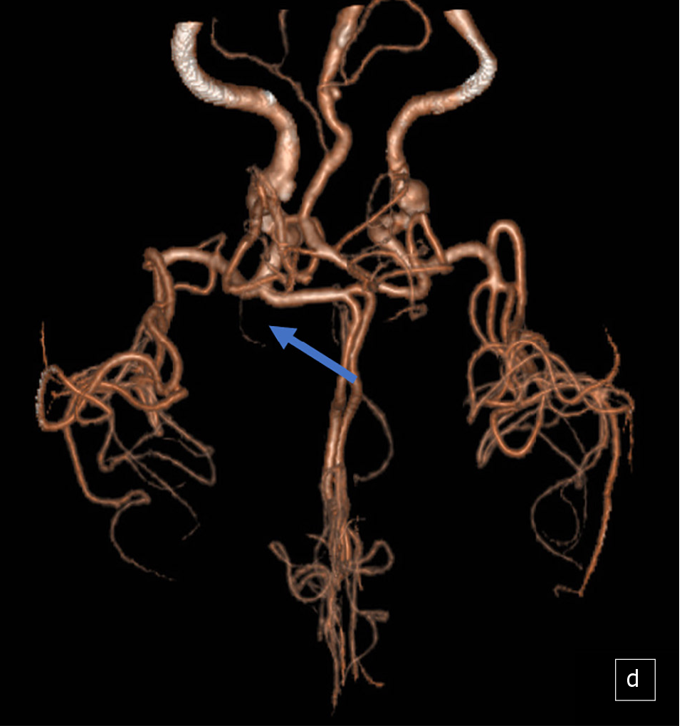
Figure 2 Computed tomography angiography examination.
This figure illustrates the location of the Percheron artery and its acute occlusion on computed tomography angiography examination.
- Citation: Nong XF, Cao X, Tan XL, Jing LY, Liu H. Percheron syndrome with memory impairment as chief manifestation: A case report. World J Clin Cases 2025; 13(13): 98937
- URL: https://www.wjgnet.com/2307-8960/full/v13/i13/98937.htm
- DOI: https://dx.doi.org/10.12998/wjcc.v13.i13.98937


