Copyright
©The Author(s) 2025.
World J Clin Cases. Jan 6, 2025; 13(1): 99746
Published online Jan 6, 2025. doi: 10.12998/wjcc.v13.i1.99746
Published online Jan 6, 2025. doi: 10.12998/wjcc.v13.i1.99746
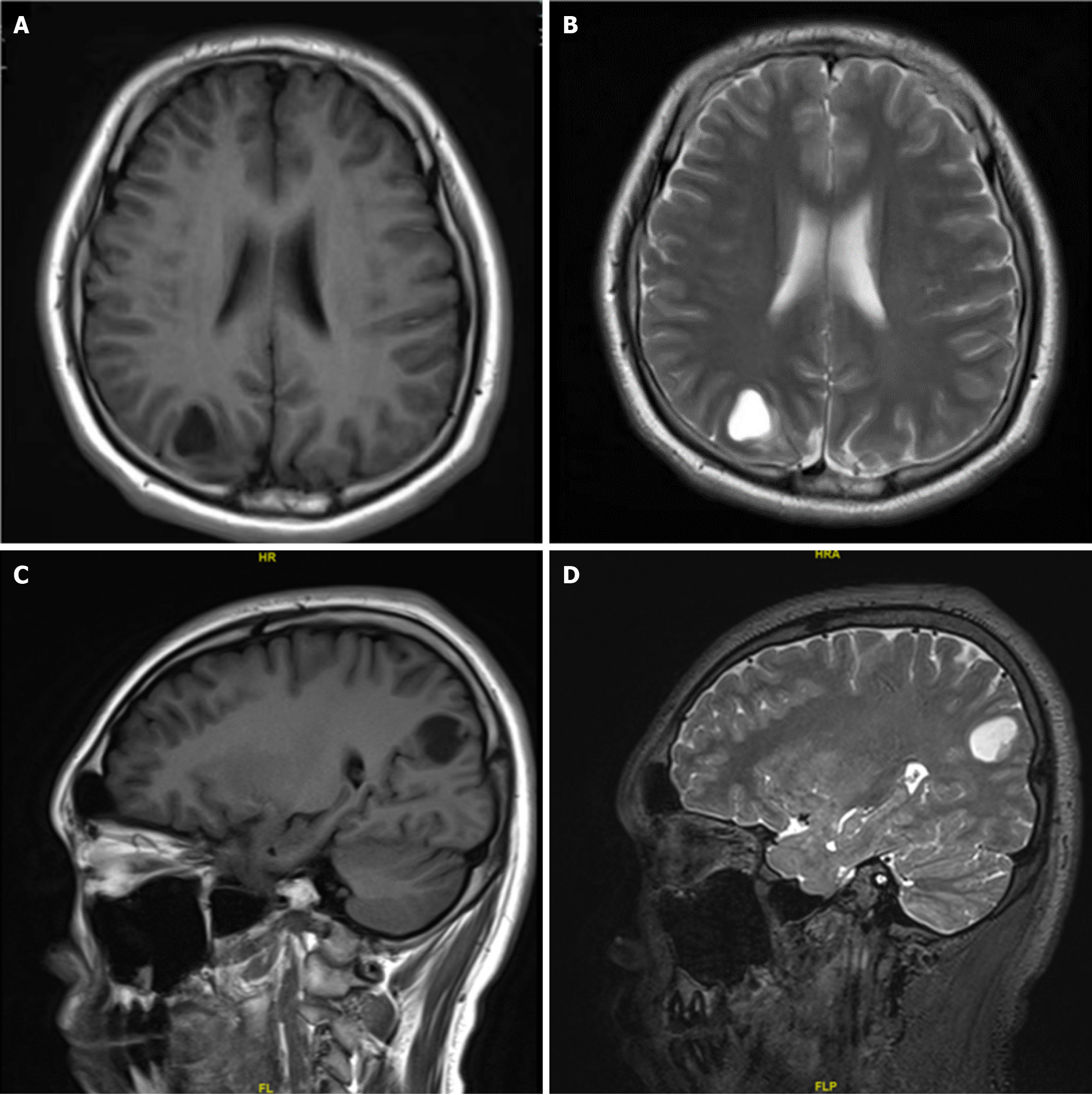
Figure 1 Magnetic resonance imaging showed a cystic mass of about 1.
9 cm × 1.5 cm × 1.9 cm under the cortex of the right parietal occipital lobe, with a uniform long T1 and long T2 signal. The cystic mass was regular, the edges were clear and smooth, and the surrounding brain tissue was slightly compressed. The remaining brain parenchyma showed no abnormal signal shadows. A: Axial T1; B: Axial T2; C: Sagittal T1; D: Sagittal T2.
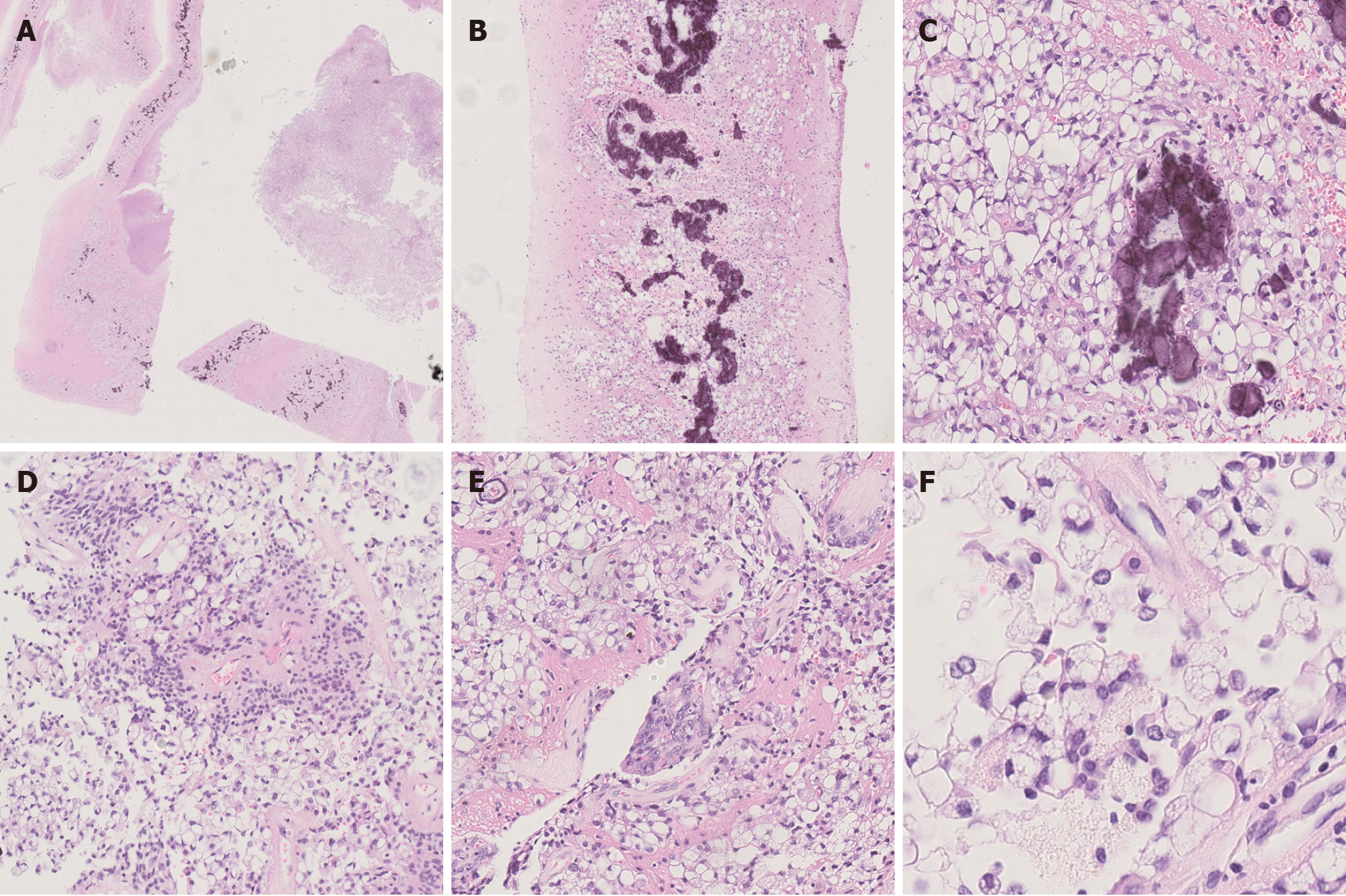
Figure 2 Histopathological findings.
A: The tissue boundary in the cystic wall-like structure was clear [hematoxylin and eosin (HE) × 40]; B: Calcifications were seen on the cyst wall (HE × 100); C: Large vacuoles were seen in the cytoplasm of tumor cells on the cyst wall (HE × 400); D: Characteristic perivascular pseudorosettes (HE × 200); E: Vascular endothelial cell proliferation was seen, resembling a glomerular structure (HE × 200); F: Vacuoles in the cytoplasm pushed the crescentic nucleus, similar to signet ring cells (HE × 1000).
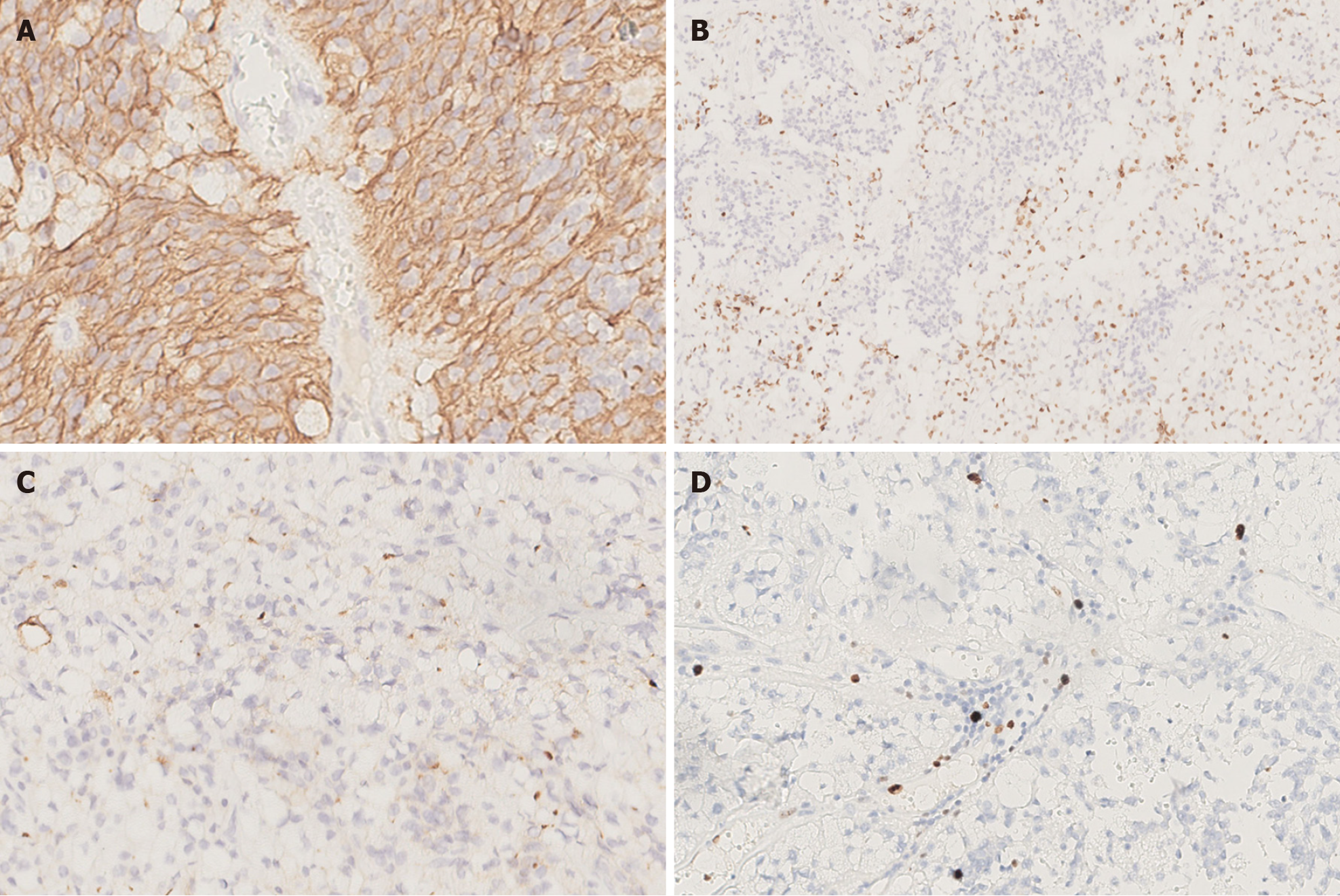
Figure 3 Immunohistochemistry findings.
A: The tumor cells were positive for GFAP [Immunohistochemistry (IHC) × 400]; B: Individual cells were positive for Olig-2 (IHC × 200); C: Significant punctate intracytoplasmic EMA immunoreactivity was observed (IHC × 400); D: The Ki-67 proliferation index was about 5% (IHC × 400).
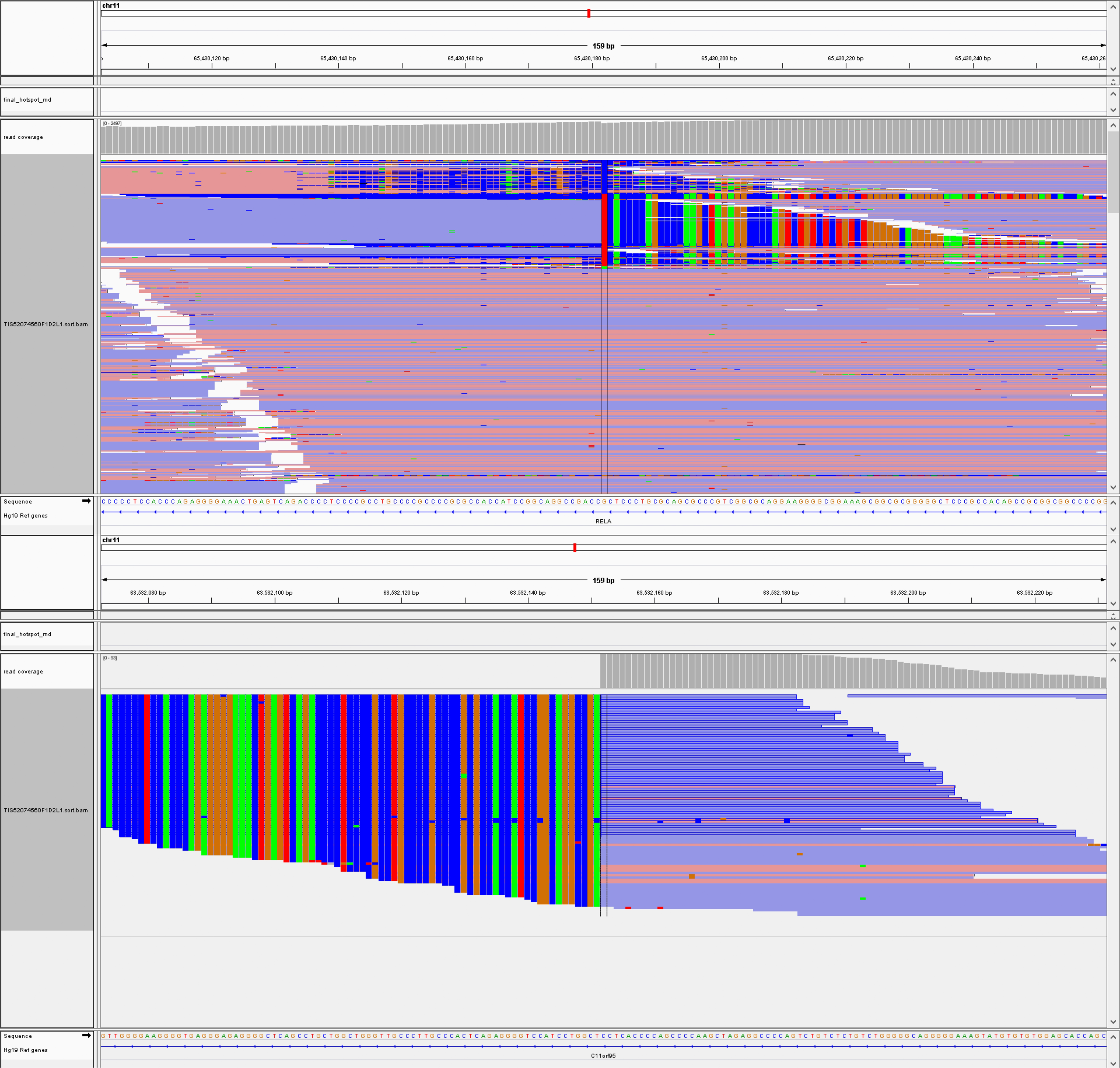
Figure 4 Genetic analysis revealed ZFTA: RELA fusion.
- Citation: Zhao XY, Yu JH, Wang YH, Liu YX, Xu L, Fu L, Yi N. Lipomatous ependymoma with ZFTA: RELA fusion-positive: A case report. World J Clin Cases 2025; 13(1): 99746
- URL: https://www.wjgnet.com/2307-8960/full/v13/i1/99746.htm
- DOI: https://dx.doi.org/10.12998/wjcc.v13.i1.99746


