Copyright
©The Author(s) 2024.
World J Clin Cases. Mar 16, 2024; 12(8): 1510-1516
Published online Mar 16, 2024. doi: 10.12998/wjcc.v12.i8.1510
Published online Mar 16, 2024. doi: 10.12998/wjcc.v12.i8.1510
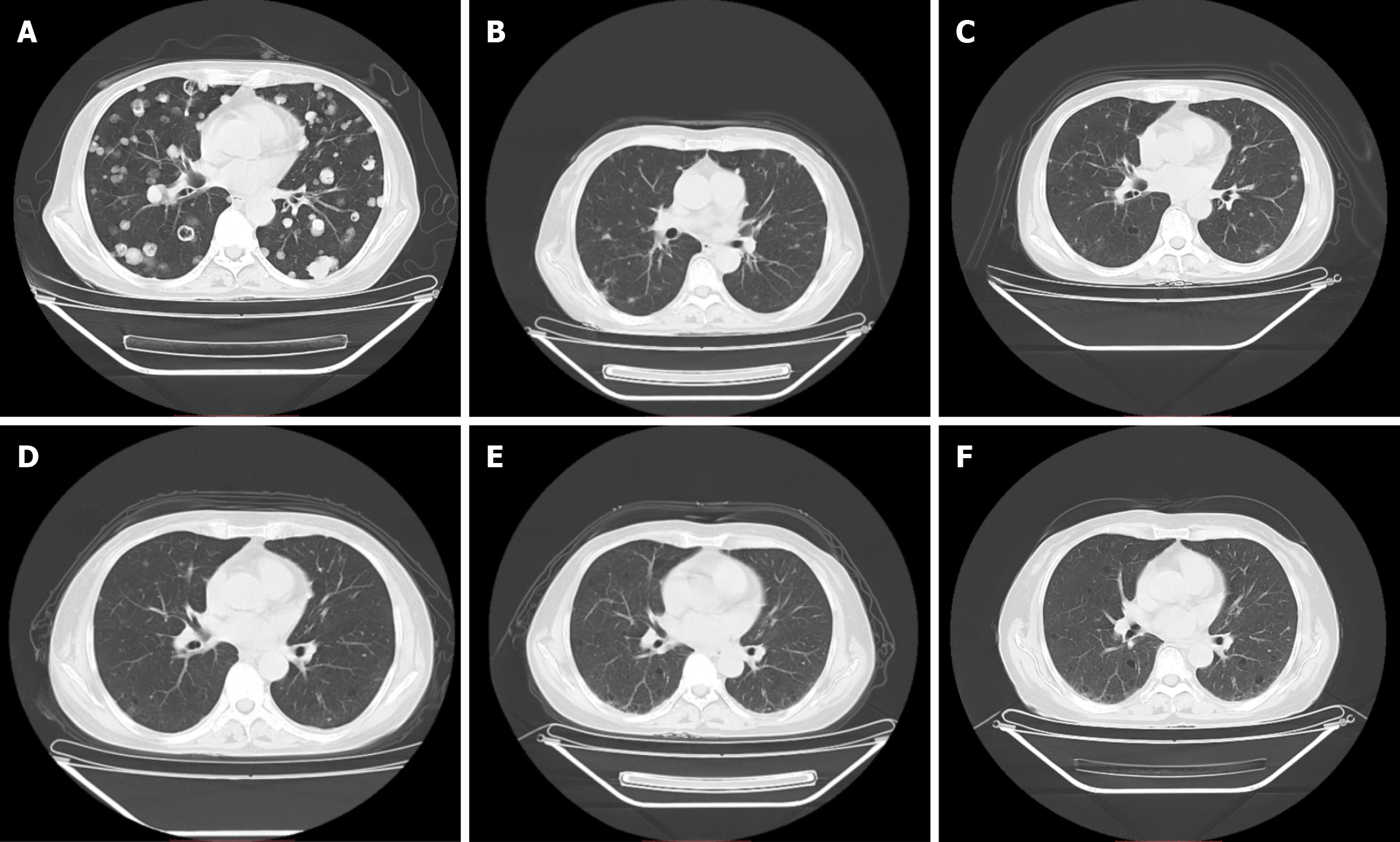
Figure 1 Computed tomography scans of the lungs were obtained before and after treatment with cadonilimab.
A: Computed tomography (CT) image of the chest on July 8, 2022, after grade 4 myelosuppression induced by chemotherapy; B: CT image of the chest on August 27, 2022, after three doses of cadonilimab; C: CT image of the chest on October 28, 2022, after treatment with cadonilimab; D: CT image of the chest on January 18, 2023, after treatment with cadonilimab; E: CT image of the chest on April 27, 2023, indicating complete remission (CR); F: CT image of the chest on July 24, 2023, showing that the patient achieved sustained CR.
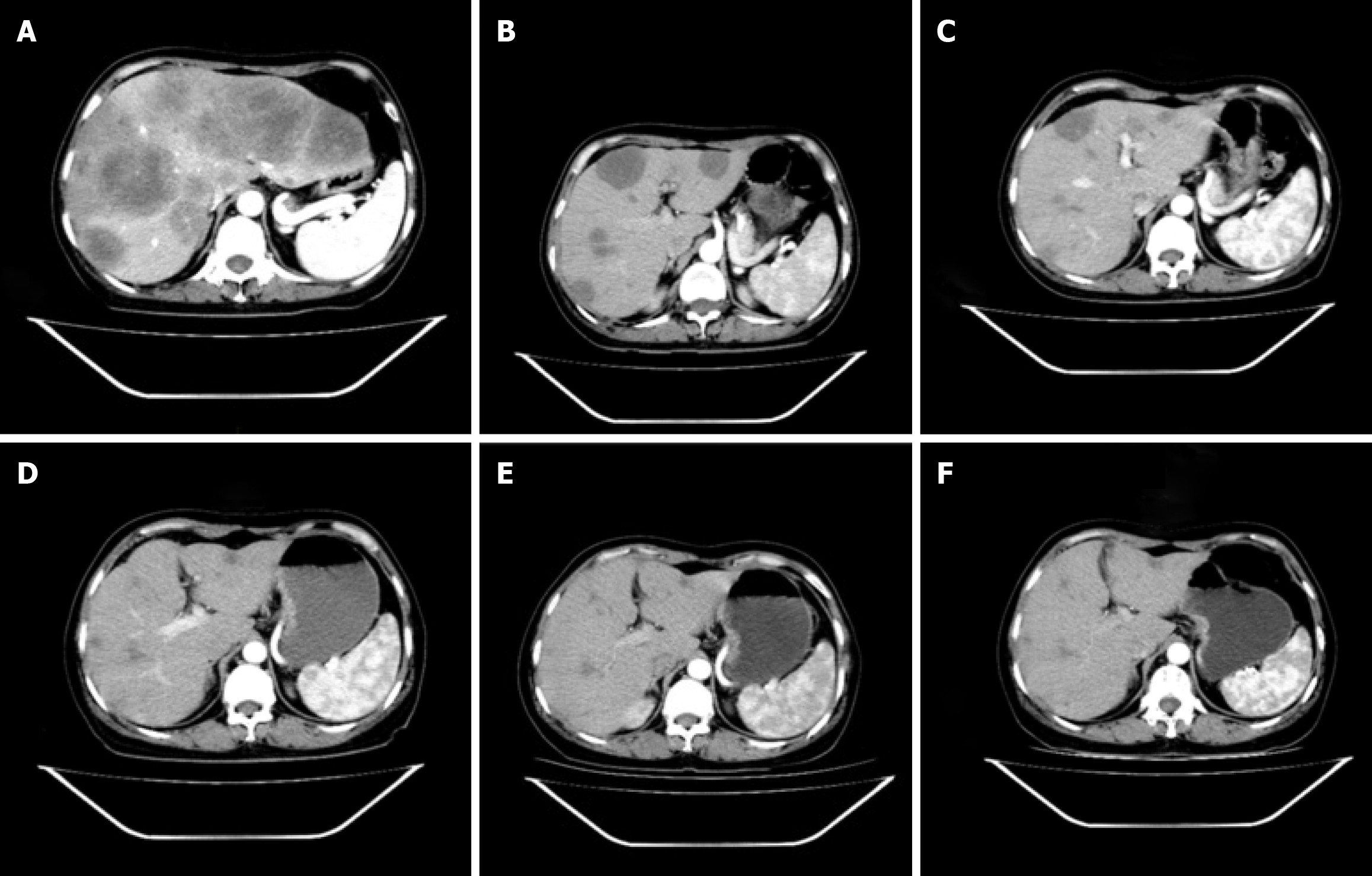
Figure 2 The arterial phase of contrast-enhanced computed tomography of the liver was conducted before and after treatment with cadonilimab.
A: Enhanced computed tomography (CT) image of the liver on July 8, 2022, after grade 4 myelosuppression was induced by chemotherapy; B: Enhanced CT image of the liver on August 27, 2022, after three doses of cadonilimab; C: Enhanced CT image of the liver on October 28, 2022, after treatment with cadonilimab; D: Enhanced CT image of the liver on January 18, 2023, after treatment with cadonilimab; E: Enhanced CT image of the liver on April 27, 2023, showing that the patient achieved complete remission (CR); F: Enhanced CT image of the liver on July 24, 2023, showing that the patient achieved sustained CR.
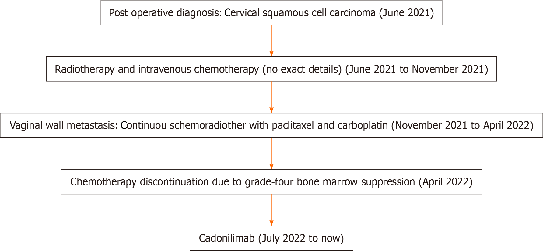
Figure 3 Critical diagnosis and treatment of the patient.
- Citation: Zhu R, Chen TZ, Sun MT, Zhu CR. Advanced cervix cancer patient with chemotherapy-induced grade IV myelosuppression achieved complete remission with cadonilimab: A case report. World J Clin Cases 2024; 12(8): 1510-1516
- URL: https://www.wjgnet.com/2307-8960/full/v12/i8/1510.htm
- DOI: https://dx.doi.org/10.12998/wjcc.v12.i8.1510


