Copyright
©The Author(s) 2024.
World J Clin Cases. Mar 16, 2024; 12(8): 1481-1486
Published online Mar 16, 2024. doi: 10.12998/wjcc.v12.i8.1481
Published online Mar 16, 2024. doi: 10.12998/wjcc.v12.i8.1481
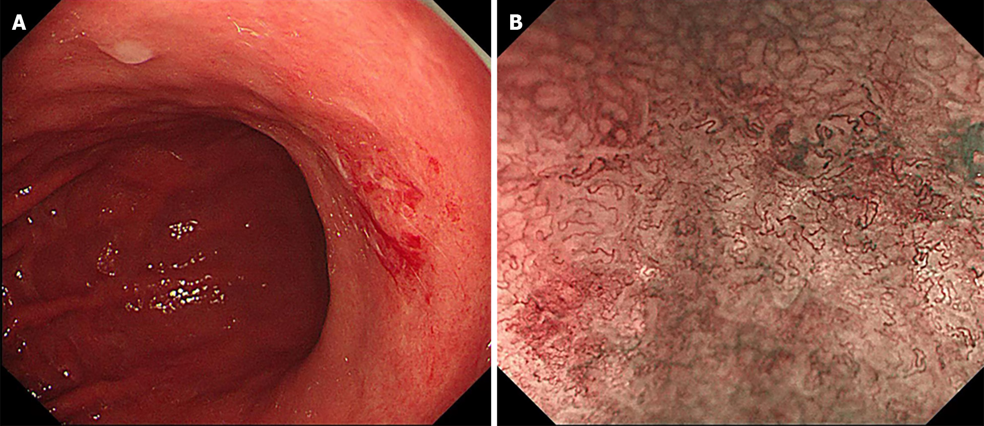
Figure 1 Gastroscopy features.
A: Endoscopy observation of the small curvature of the stomach; B: Magnified Narrow band imaging examination of the gastric mucosa. NBI: Narrow band imaging.
Figure 2 Confocal endomicroscopy features.
A and B: Differentiated adenocarcinoma displaying a disorganized epithelium with dark and irregular glands; C: Undifferentiated adenocarcinoma characterized by dark and irregular cells with no identifiable glandular structures.
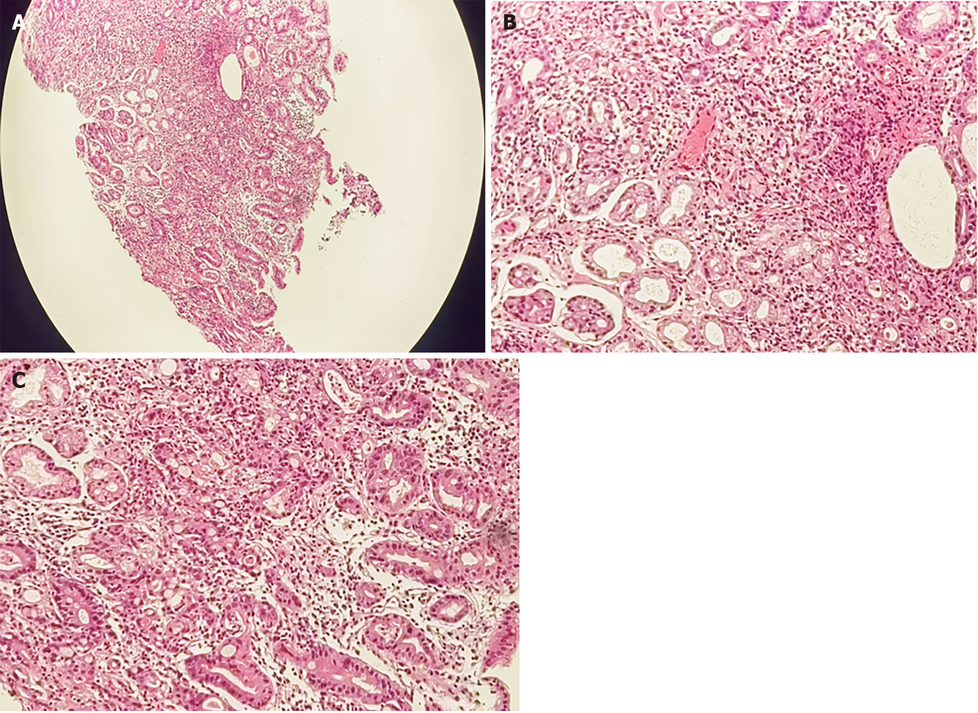
Figure 3 Histological examinations of the gastric mucosa by Hematoxylin-eosin staining revealed the pathology.
A: Under 40 × magnification; B and C: Under 200 × magnification.
- Citation: Lou JX, Wu Y, Huhe M, Zhang JJ, Jia DW, Jiang ZY. Diagnosis of poorly differentiated adenocarcinoma of the stomach by confocal laser endomicroscopy: A case report. World J Clin Cases 2024; 12(8): 1481-1486
- URL: https://www.wjgnet.com/2307-8960/full/v12/i8/1481.htm
- DOI: https://dx.doi.org/10.12998/wjcc.v12.i8.1481


