Copyright
©The Author(s) 2024.
World J Clin Cases. Feb 26, 2024; 12(6): 1144-1149
Published online Feb 26, 2024. doi: 10.12998/wjcc.v12.i6.1144
Published online Feb 26, 2024. doi: 10.12998/wjcc.v12.i6.1144
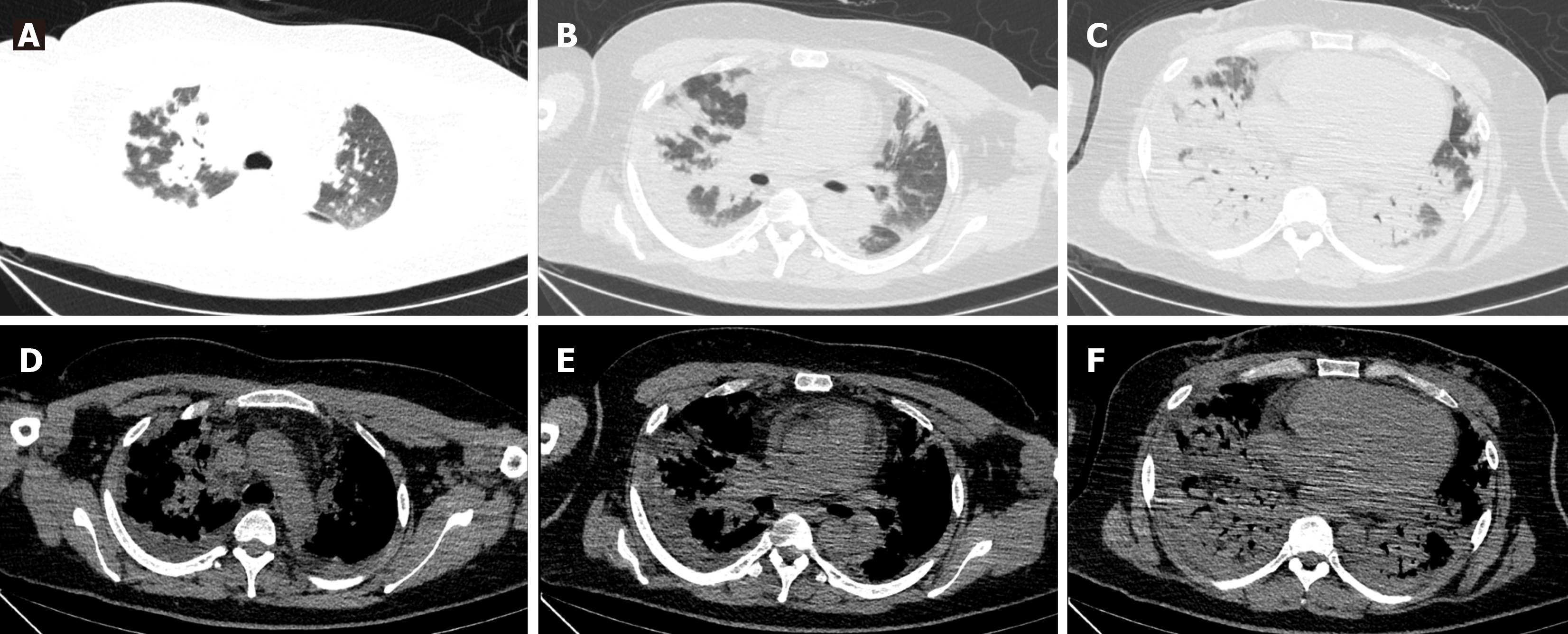
Figure 1 Lung computed tomography examination in admission.
There were diffuse multiple patchy heightening shadows in both lungs, with a partial bronchial inflation sign. A: Both upper lungs in the lung window; B: Middle lobe and tongue lobe in the lung window; C: Both lower lungs in the lung window; D: Both upper lungs in the mediastinal window; E: Middle lobe and tongue lobe in the mediastinal window; F: Both lower lungs in the mediastinal window.
Figure 2 Lung computed tomography examination after methylprednisolone 40 mg paxlovid 6 d.
Diffuse multiple patches of increased density in both upper lungs were significantly absorbed. A: Both upper lungs in the lung window; B: Middle lobe and tongue lobe in the lung window; C: Both lower lungs in the lung window.
Figure 3 Color ultrasound-guided percutaneous lung puncture.
Organizing pneumonia with alveolar septal thickening was found under the microscope (hematoxylin-eosin). A: Original magnification × 10; B: Original magnification × 20; C: Original magnification × 40.
Figure 4 Lung computed tomography examination at 1 mo follow-up.
Diffuse multiple consolidations in both lower lungs were significantly absorbed. A: Both upper lungs in the lung window; B: Middle lobe and tongue lobe in the lung window; C: Both lower lungs in the lung window.
- Citation: Wu XX, Cui J, Wang SY, Zhao TT, Yuan YF, Yang L, Zuo W, Liao WJ. Clinical evolution of antisynthetase syndrome-associated interstitial lung disease after COVID-19 in a man with Klinefelter syndrome: A case report. World J Clin Cases 2024; 12(6): 1144-1149
- URL: https://www.wjgnet.com/2307-8960/full/v12/i6/1144.htm
- DOI: https://dx.doi.org/10.12998/wjcc.v12.i6.1144


