Copyright
©The Author(s) 2024.
World J Clin Cases. Feb 16, 2024; 12(5): 1004-1009
Published online Feb 16, 2024. doi: 10.12998/wjcc.v12.i5.1004
Published online Feb 16, 2024. doi: 10.12998/wjcc.v12.i5.1004
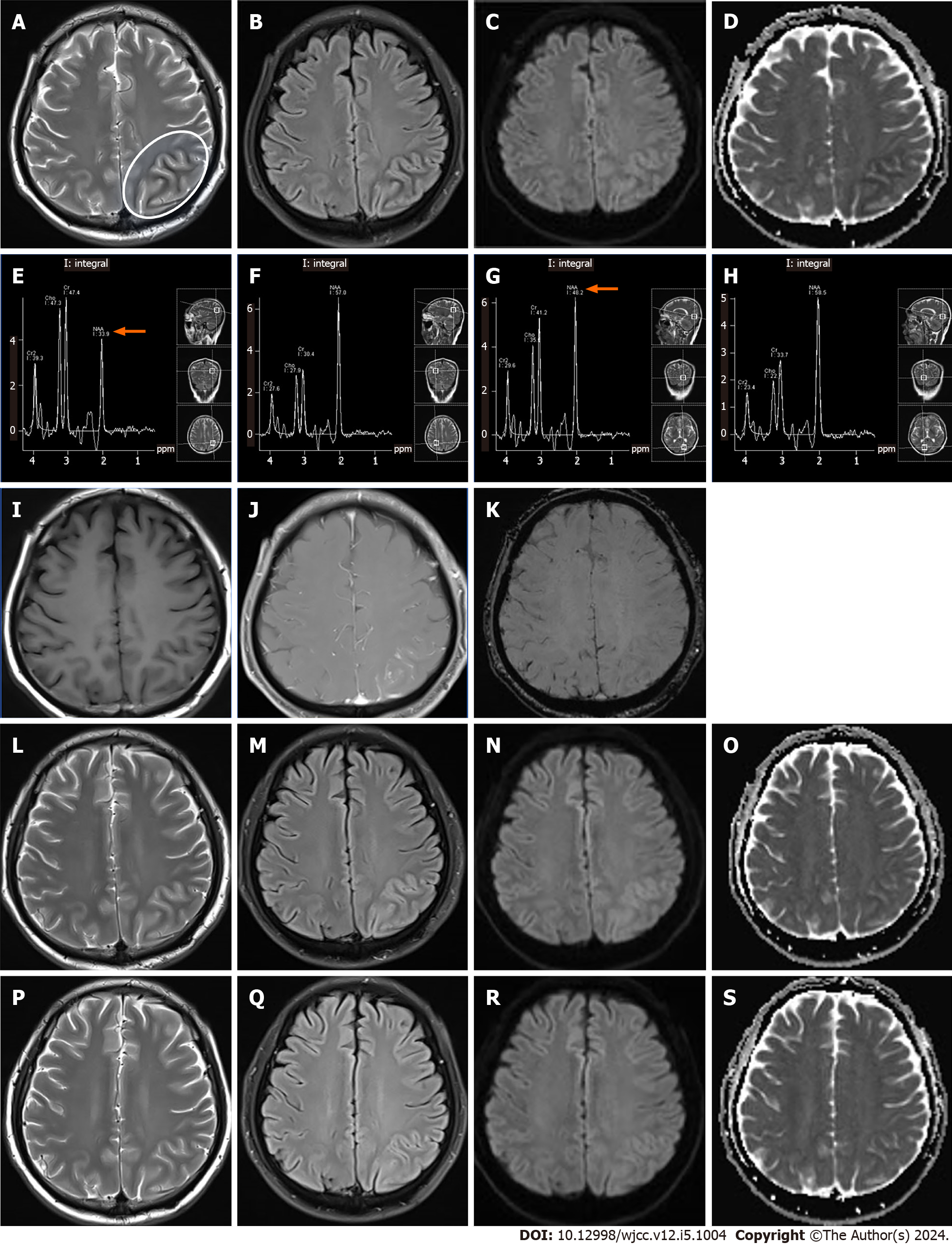
Figure 1 Head magnetic resonance imaging findings of a patient with non-ketotic hyperglycaemic seizures with hyperhomocysteinaemia.
A-D: Images at admission, showing the left parieto-occipital cortex (white arrow) on (A) T2-weighted images (T2WI) and (B) fluid-attenuated inversion recovery (FLAIR) sequence with cortical swelling and hyperintense T2 signal. (C) Diffusion-weighted imaging (DWI) shows restricted diffusion in the left parieto-occipital cortex and (D) low signal in the apparent diffusion coefficient (ADC) map; E–H: Magnetic resonance spectroscopy (MRS) images on the day after admission, showing (E) decreased N-acetylaspartate (NAA) peaks (white arrows) in the left parieto-occipital subcortex, increased creatine (Cr) and choline (Cho) peak, and (F) no significant alterations in the right parieto-occipital subcortex. MRS also showed (G) decreased NAA peaks (white arrows) in the left parieto-occipital cortex, and increased Cr and Cho peaks, less evident than in the corresponding subcortex in panel (E). (H) The right parieto-occipital cortex showed no significant alterations; I–K: Contrast-enhanced magnetic resonance imaging (MRI) images on the day after admission, showing no significant alterations on (E) T1-weighted images, with swelling of the left parieto-occipital gyrus and localized soft meningeal enhancement in the parietal lobe (J). No significant alterations were noted on susceptibility-weighted imaging (K); L–O: MRI findings after 12 d of treatment, showing alleviation of the subcortical hypointensities on (L) T2WI and (M) FLAIR images, with more pronounced hyperintensity in the left parieto-occipital cortex in the latter than at admission. (N) DWI and (O) ADC map showed no significant changes; P–S: Follow-up MRI images 20 d after discharge, showing normalisation of the left parieto-occipital cortex and subcortical areas on (P) T2WI and (Q) FLAIR images. (R) DWI and (S) ADC map showed no significant alterations.
- Citation: Wu J, Feng H, Zhao Y, Li J, Li T, Li K. Neuroimaging features in a patient with non-ketotic hyperglycaemic seizures: A case report. World J Clin Cases 2024; 12(5): 1004-1009
- URL: https://www.wjgnet.com/2307-8960/full/v12/i5/1004.htm
- DOI: https://dx.doi.org/10.12998/wjcc.v12.i5.1004


