Copyright
©The Author(s) 2024.
World J Clin Cases. Jan 26, 2024; 12(3): 657-664
Published online Jan 26, 2024. doi: 10.12998/wjcc.v12.i3.657
Published online Jan 26, 2024. doi: 10.12998/wjcc.v12.i3.657
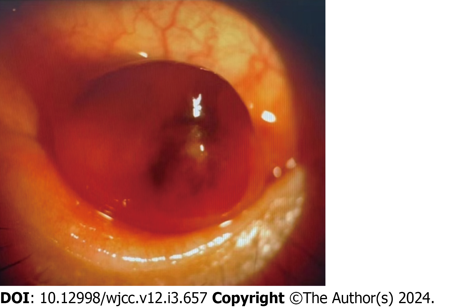
Figure 1 Anterior segment image showed an 8 mm slightly elevated, sessile, pink-colored sub-epithelial mass described as a “salmon patch” in the conjunctival sac of the left eye.
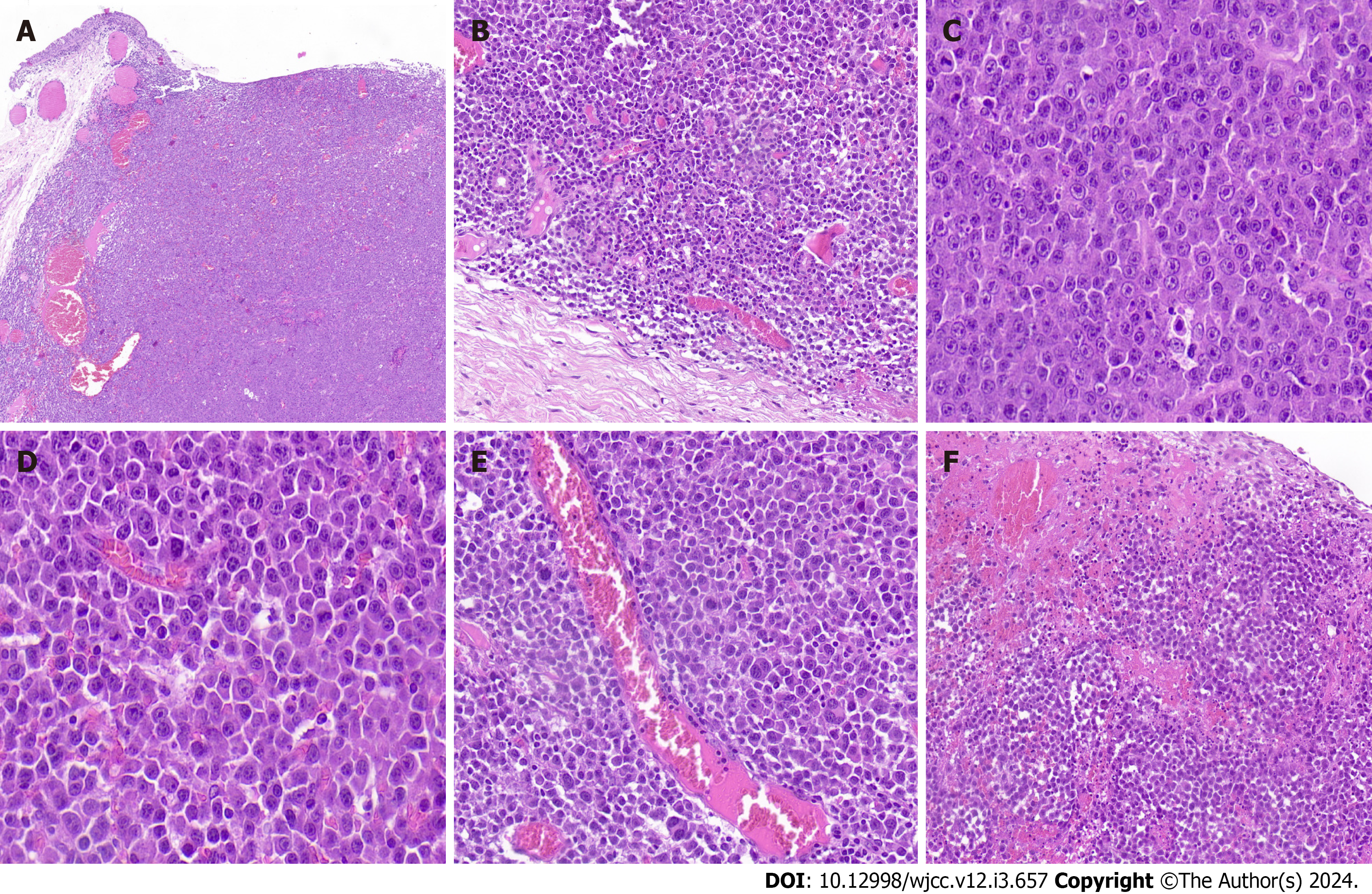
Figure 2 Anaplastic lymphoma kinase-positive large B-cell lymphoma.
A: The tumor was well-demarcated and showed a diffusely infiltrate of monomorphicatypical lymphoid cells in the stroma of the conjunctival subepithelial [hematoxylin and eosin (HE) ×100]; B: The accessory lacrimal glands were involved (HE × 400); C: The lymphoid cells displayed immunoblastic morphology (HE × 1000); D: The lymphoid cells with plasmablastic morphology (HE × 1000); E: the pleomorphic neoplastic giant cells were present focally (HE × 400); F: Necrosis, abundant apoptotic debris and haemorrhage were observed (HE × 400).
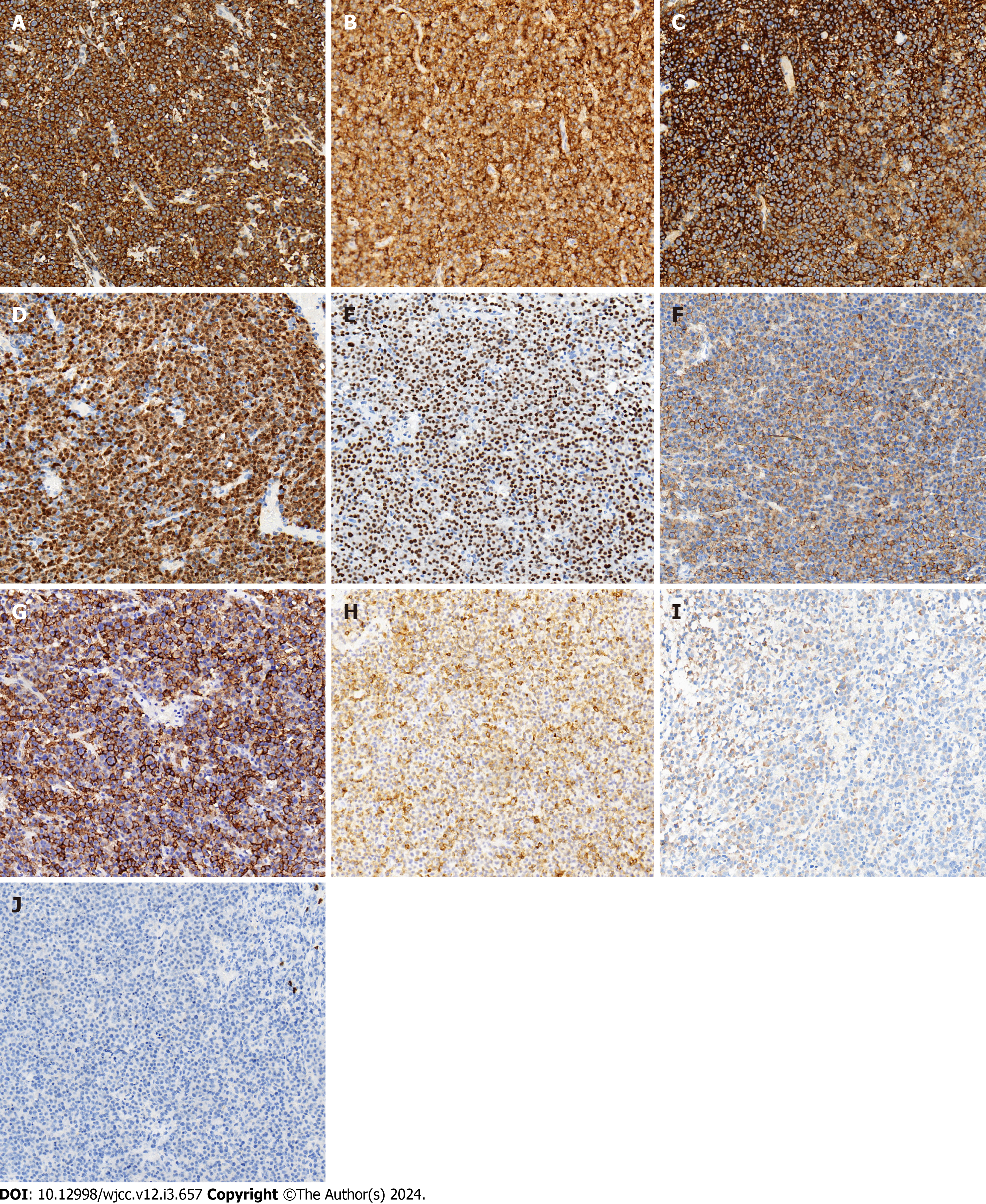
Figure 3 Immunohistochemical stains of anaplastic lymphoma kinase-positive large B-cell lymphoma (× 400).
A-G: The large atypical cells were diffusely positive for anaplastic lymphoma kinase with a characteristic cytoplasmic granular staining pattern (A), CD10 (B), CD138 (C), BOB.1 (D), OCT-2 (E), CD4 (F) and EMA (G); H-J: Focally positive for CD79a (H) and IgA (I); negative for CD20 (J).
- Citation: Guo XH, Li CB, Cao HH, Yang GY. Primary anaplastic lymphoma kinase-positive large B-cell lymphoma of the left bulbar conjunctiva: A case report. World J Clin Cases 2024; 12(3): 657-664
- URL: https://www.wjgnet.com/2307-8960/full/v12/i3/657.htm
- DOI: https://dx.doi.org/10.12998/wjcc.v12.i3.657


