Copyright
©The Author(s) 2024.
World J Clin Cases. Jan 26, 2024; 12(3): 575-581
Published online Jan 26, 2024. doi: 10.12998/wjcc.v12.i3.575
Published online Jan 26, 2024. doi: 10.12998/wjcc.v12.i3.575
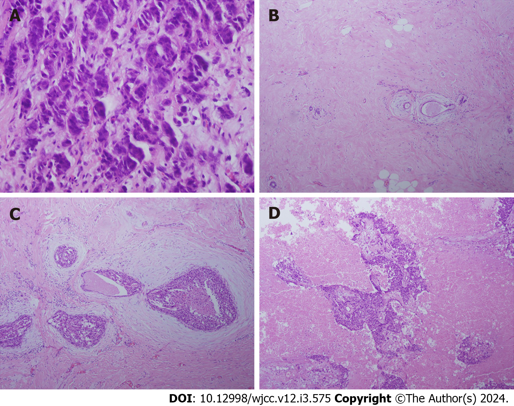
Figure 1 High power microscopic view of lesion tissue.
A: Aspiration biopsy tissue of the ipsilateral breast (40 ×); B: Lesion tissue from modified radical mastectomy after preoperative neoadjuvant treatment (10 ×); C: Lesion tissue from modified radical mastectomy after preoperative neoadjuvant treatment (20 ×); D: Lesion tissue from resected brain metastasis (20 ×).
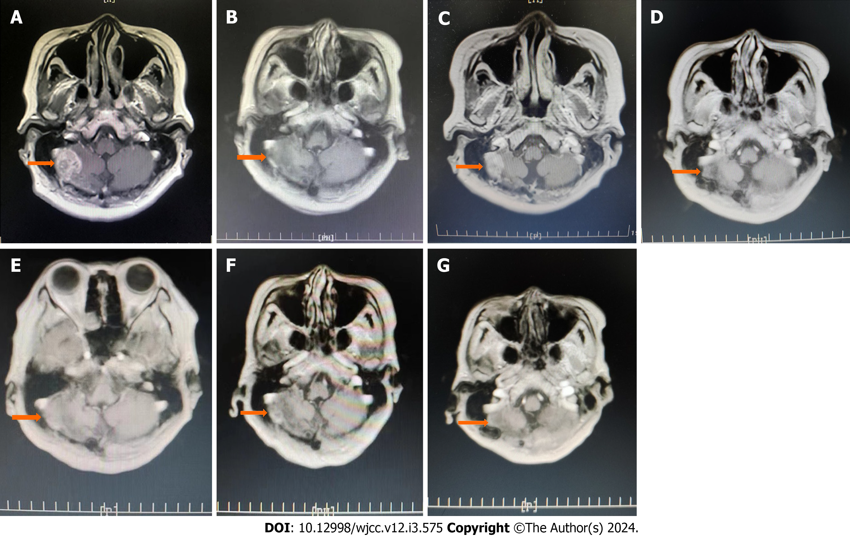
Figure 2 Magnetic resonance imaging.
A: Recurrence after brain metastasis resection, tumor size, 25 mm × 20 mm (October 29, 2021); B: After 10 cycles of systemic treatment, the tumor shrank considerably (January 26, 2021); C: After 16 cycles of treatment, before 6 cycles of radiation therapy, the tumor size was approximately 27 mm × 18 mm (October 8, 2021); D: The tumor size was approximately 9 mm × 7 mm (May 7, 2022); E: After 10 cycles of treatment, the size of the tumor had not changed notably, i.e., approximately 10 mm × 8 mm (August 1, 2022); F: After 19 cycles of treatment, the size of the tumor was approximately 11 mm × 10 mm (February 8, 2023); G: After 31 cycles of treatment, the tumor size was approximately 11 mm × 9 mm (April 2, 2023).
- Citation: Dou QQ, Sun TT, Wang GQ, Tong WB. Inetetamab combined with pyrotinib and chemotherapy in the treatment of breast cancer brain metastasis: A case report. World J Clin Cases 2024; 12(3): 575-581
- URL: https://www.wjgnet.com/2307-8960/full/v12/i3/575.htm
- DOI: https://dx.doi.org/10.12998/wjcc.v12.i3.575


