Copyright
©The Author(s) 2024.
World J Clin Cases. Sep 26, 2024; 12(27): 6105-6110
Published online Sep 26, 2024. doi: 10.12998/wjcc.v12.i27.6105
Published online Sep 26, 2024. doi: 10.12998/wjcc.v12.i27.6105
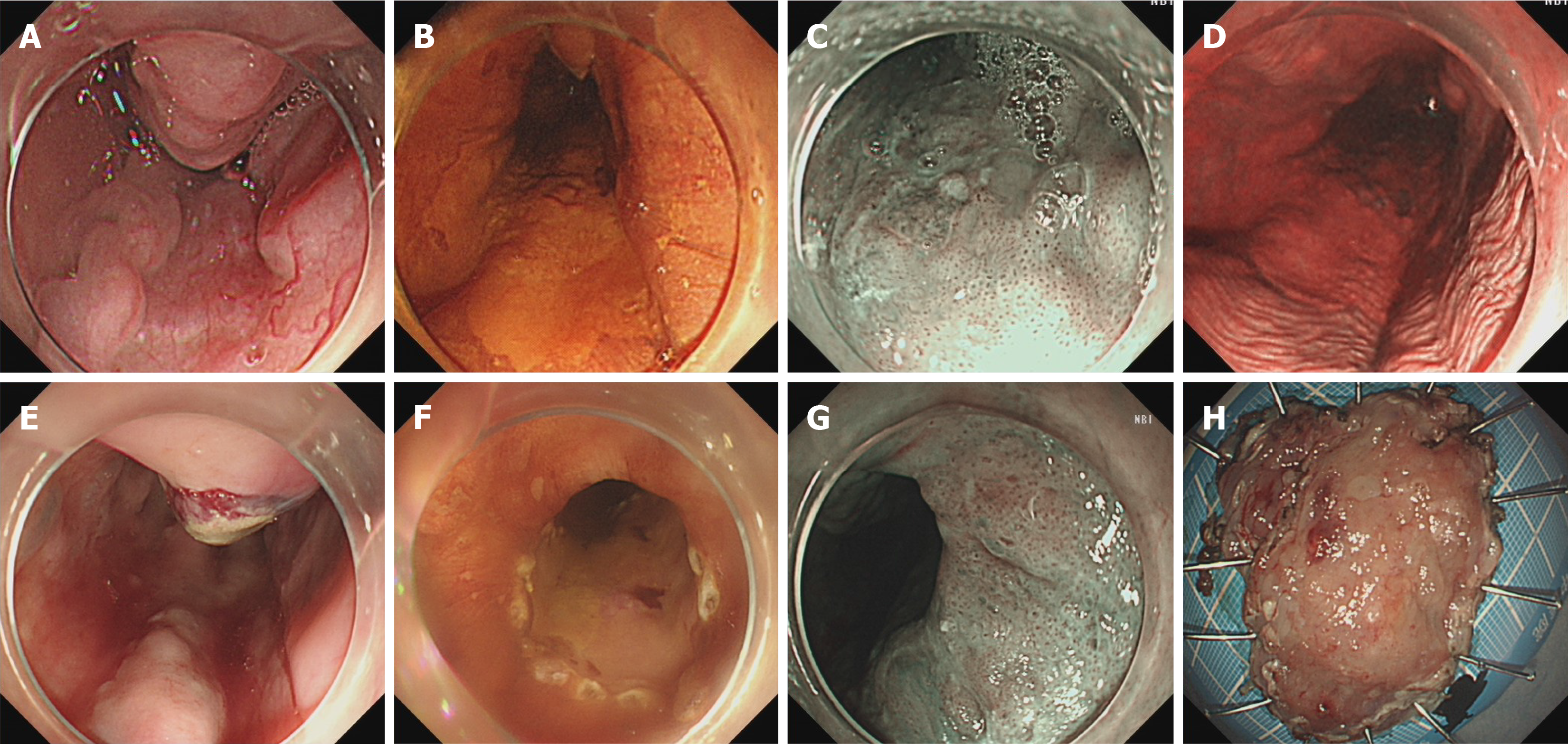
Figure 1 Endoscopic images.
A: Three esophageal varices with a diameter of 0.6 cm; B: IIb-type lesion with a pink color that could not be stained by iodine; C: Irregular blood vessels on narrow-band imaging-magnetic endoscopy (NBI-ME); D: Tatami sign; E: Gum discharged after endoscopic selective variceal debridement and endoscopic injection sclerotherapy; F: Demarcation line and marker; G: Irregular blood vasculature on NBI-ME; H: Postoperative specimen.
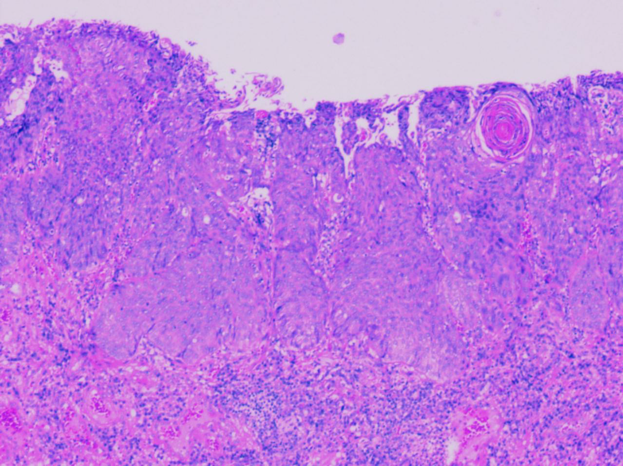
Figure 2
Pathology revealed 0-IIa + IIb type squamous cell carcinoma with moderate differentiation.
Figure 3 Timeline.
ESVD: Esophageal solitary venous dilatation; EIS: Endoscopic injection sclerotherapy; ESD: Endoscopic submucosal dissection.
- Citation: Xu L, Chen SS, Yang C, Cao HJ. Successful endoscopic treatment of superficial esophageal cancer in a patient with esophageal variceal bleeding: A case report. World J Clin Cases 2024; 12(27): 6105-6110
- URL: https://www.wjgnet.com/2307-8960/full/v12/i27/6105.htm
- DOI: https://dx.doi.org/10.12998/wjcc.v12.i27.6105


