Copyright
©The Author(s) 2024.
World J Clin Cases. Sep 16, 2024; 12(26): 5885-5892
Published online Sep 16, 2024. doi: 10.12998/wjcc.v12.i26.5885
Published online Sep 16, 2024. doi: 10.12998/wjcc.v12.i26.5885
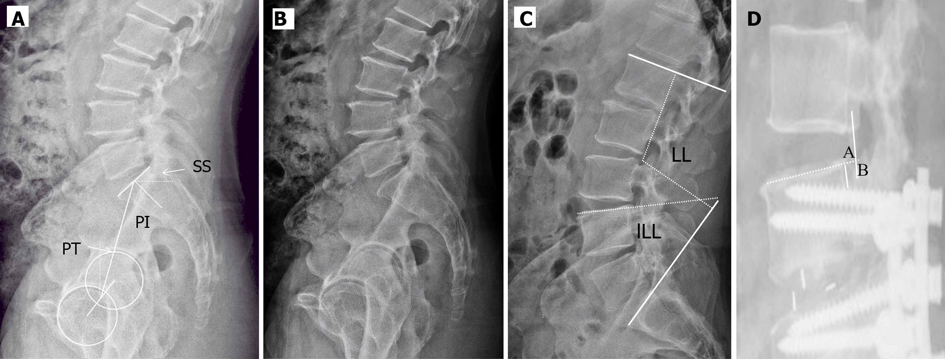
Figure 1 Pelvic and spinal parameters and retrograde distance of cranial adjacent segments.
A: Pelvic incidence: The angle between the line perpendicular to the sacral plate at its midpoint and the line connecting this point to the axis of the femoral heads; Pelvic tilt: The angle between the line connecting the midpoint of the sacral plate to the femoral head axis and the vertical axis; Sacral slope: The angle between the superior plate of S1 and a horizontal line; B: Lumbar lordosis (LL): The angle between the superior endplate of the L1 vertebra and the superior endplate of the S1; Lower LL: The angle between the superior endplate of the L4 vertebra and the superior endplate of the S1; C: Retrograde distance of cranial adjacent segments: The distance between point A and point B; point A: The dorsal edges of the cranial endplate of the inferior vertebral, point B: The intersection point between the line crossing the dorsal edges of the caudal endplate of the superior vertebral and perpendicular to the cranial endplate of the inferior vertebral. PI: Pelvic incidence; PT: Pelvic tilt; LL: Lumbar lordosis; lLL: Lower LL; SS: Sacral slope.
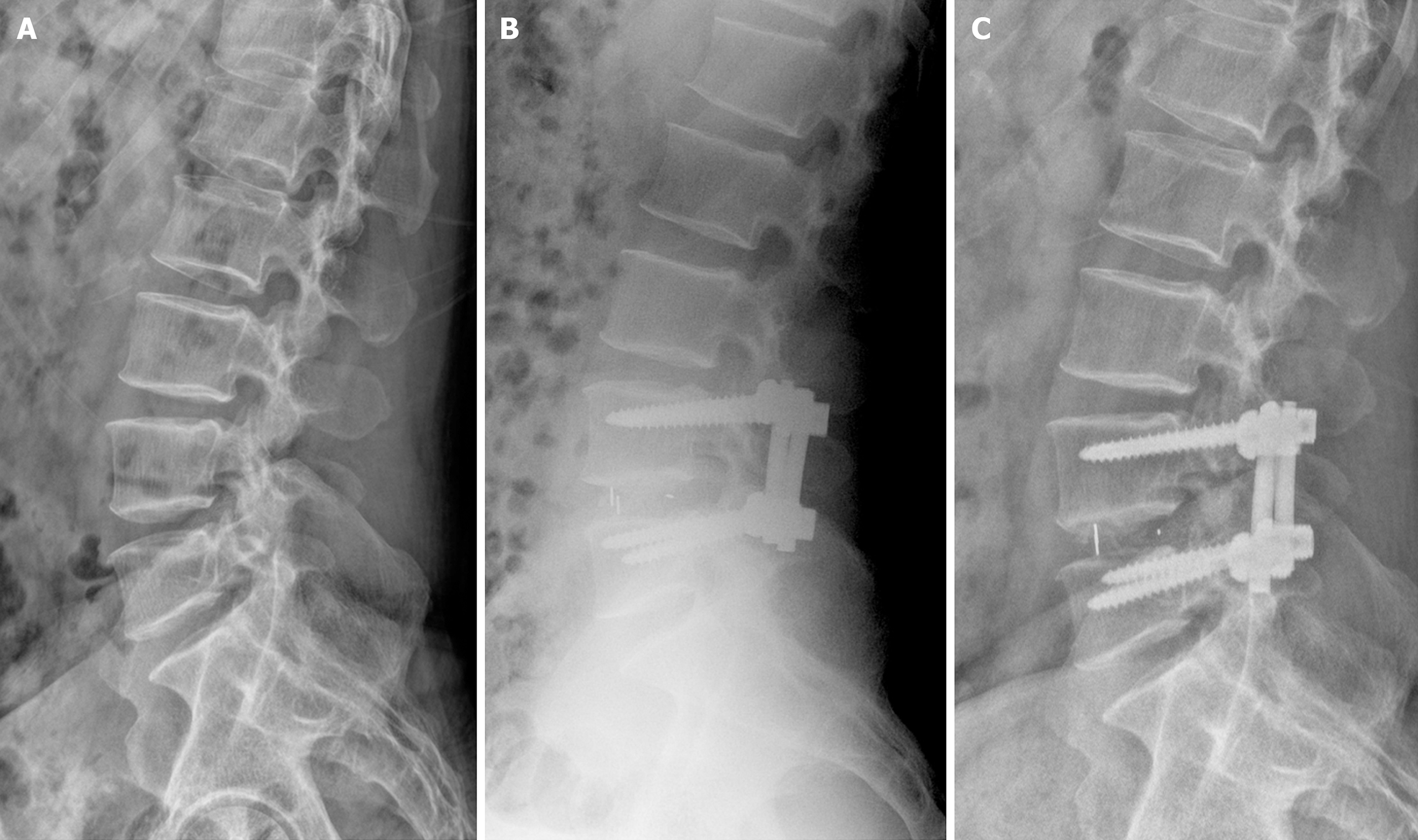
Figure 2 X-rays of a patient who underwent transforaminal lumbar interbody fusion surgery.
A: Male, 51 years, lumbar disc herniation; B: postoperative X-ray: Pelvic incidence (PI) 57°, lumbar lordosis (LL) 59°, Lower lumbar lordosis 32°, |PI-LL| 2°, lordosis distribution index 54.2%; C: 27 months after the operation: Retrograde distance of cranial adjacent segments 4mm.
- Citation: Zhu JJ, Wang Y, Zheng J, Du SY, Cao L, Yang YM, Zhang QX, Xie DD. Risk factors and clinical significance of posterior slip of the proximal vertebral body after lower lumbar fusion. World J Clin Cases 2024; 12(26): 5885-5892
- URL: https://www.wjgnet.com/2307-8960/full/v12/i26/5885.htm
- DOI: https://dx.doi.org/10.12998/wjcc.v12.i26.5885


