Copyright
©The Author(s) 2024.
World J Clin Cases. Aug 16, 2024; 12(23): 5431-5440
Published online Aug 16, 2024. doi: 10.12998/wjcc.v12.i23.5431
Published online Aug 16, 2024. doi: 10.12998/wjcc.v12.i23.5431
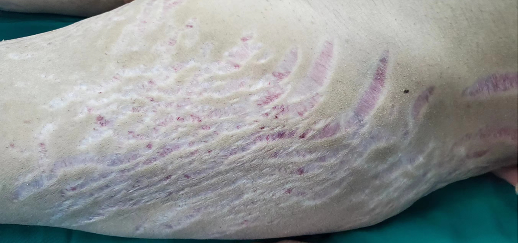
Figure 1 Physical examination.
The 18-year-old patient with purple stripes on his outer thigh.
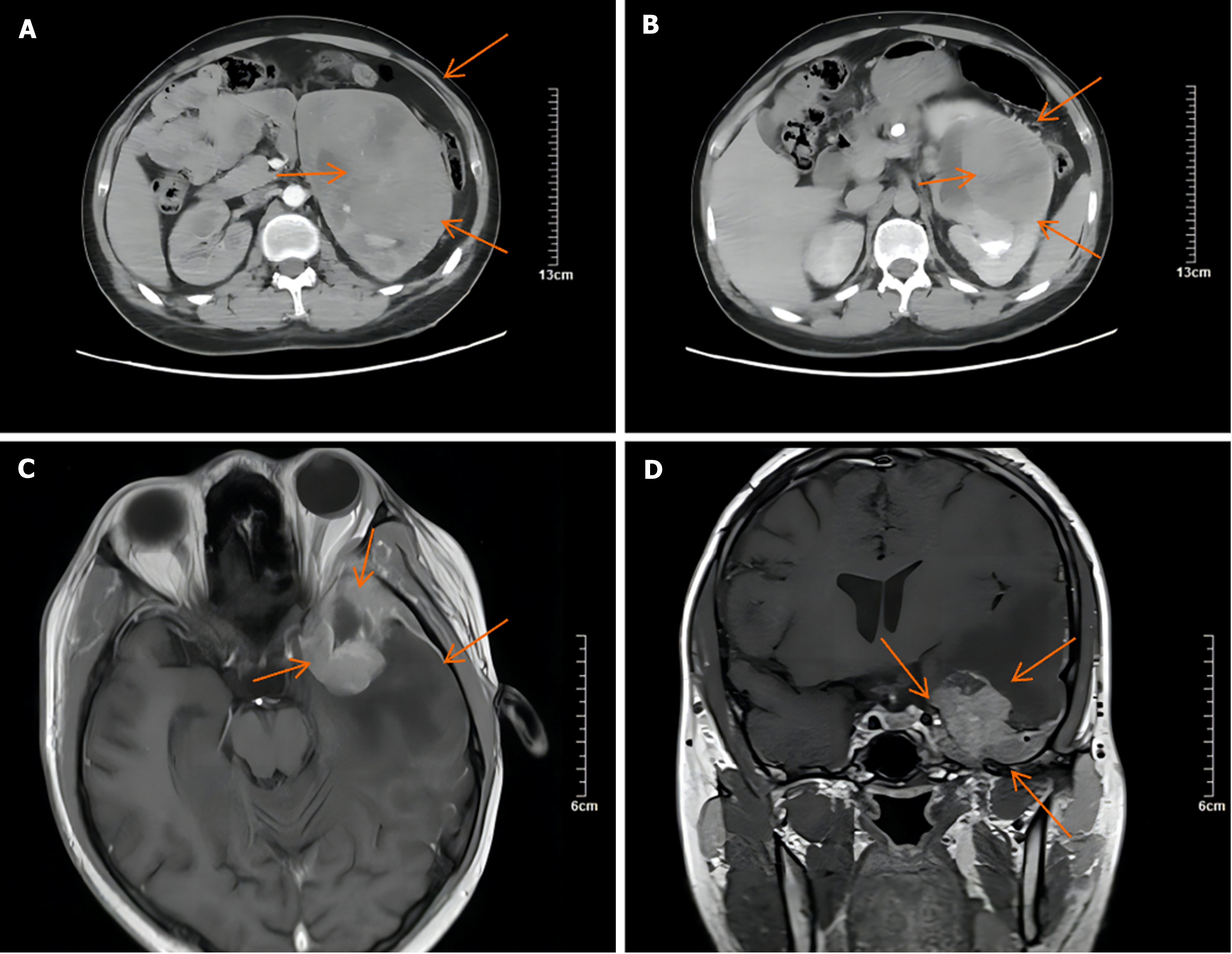
Figure 2 Computed tomography and magnetic resonance imaging.
A and B: Corticomedullary and excretory phase axial computed tomography shows that the large tumor (11.65 cm × 10.91 cm) showed scattered irregular enhancement with large areas of necrosis; C and D: Magnetic resonance imaging coronal and axial view of the head showed mixed signals in the left temporal lobe, suggesting tumor metastasis.
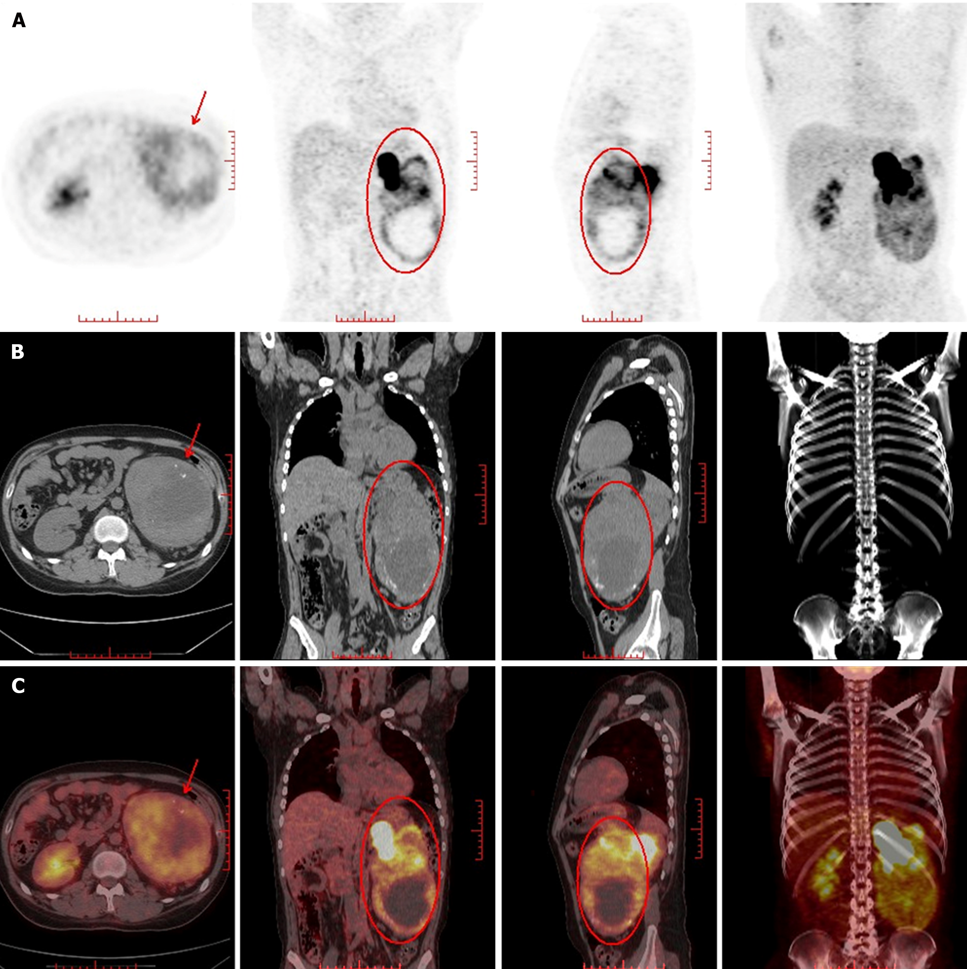
Figure 3 Nuclear medicine image of the primary tumor.
A: Increased radioactivity in the left kidney; B: A large mass of mixed density was found in the left kidney. The large cross section of the lesion was about 11.8 cm × 9.7 cm; C: The solid component showed uneven radioactive concentration, Maximum Standardized Uptake Value 10.4, the left side of the kidney was compressed and displaced upward, and the renal calyx was dilated and hydronephrosis.
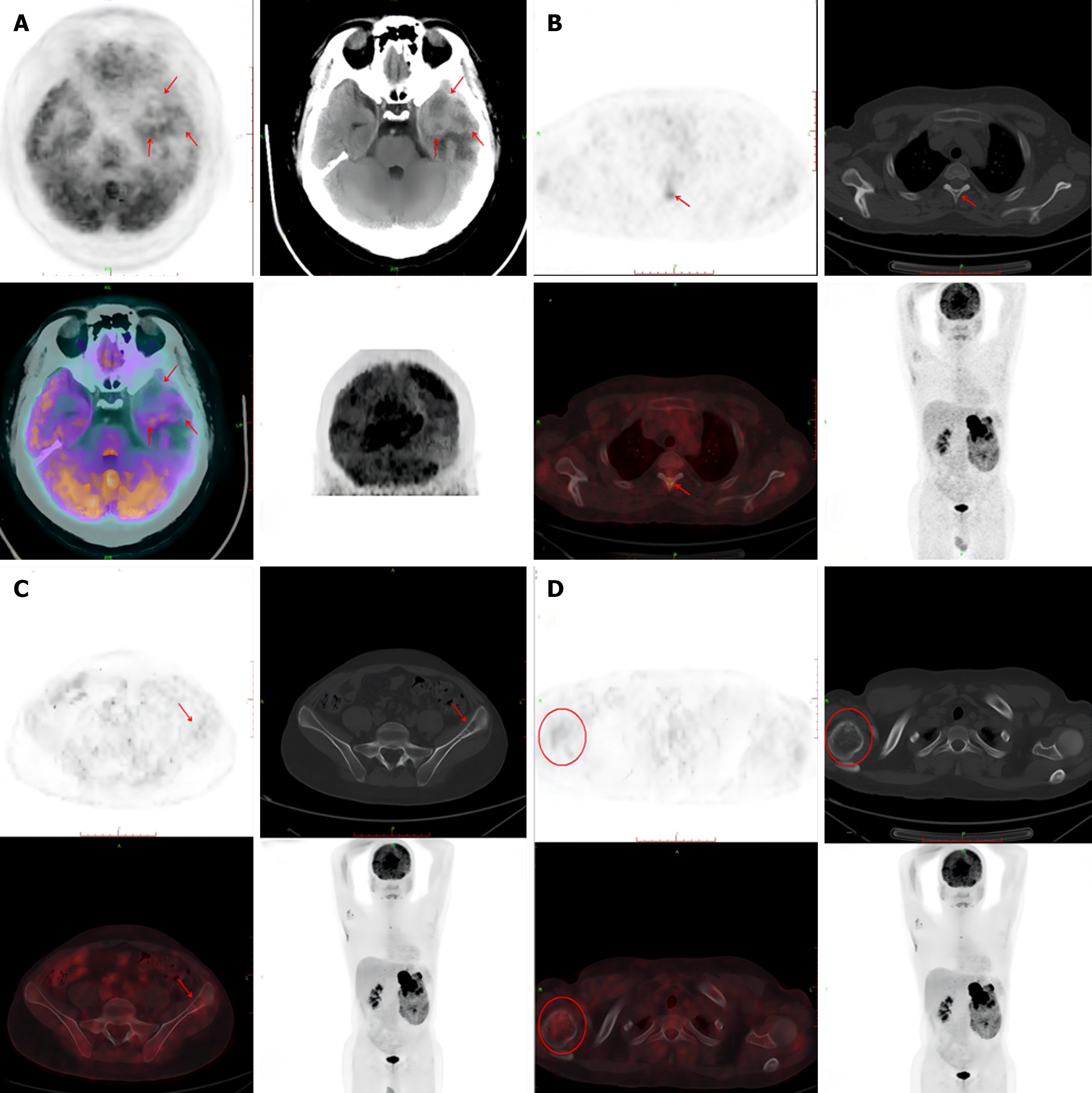
Figure 4 Nuclear medicine image of metastatic tumors.
A: The shape of the brain was as usual, with a slightly high-density mass in the left temporal pole and a few low-density shadows in the center. The larger section was about 3.3 cm × 2.2 cm. The periphery of the lesion showed large patches of low-density edema. PET showed annular high uptake of radioactivity in the lesion, Maximum Standardized Uptake Value (SUVmax) 9.1; B: High radioactive uptake shadow was found in the thoracic third vertebral arch plate, SUVmax 5.0, and no obvious bone destruction was found in the corresponding part; C and D: Left iliac wing, right humeral head and local bone of density is decreas, and a small amount of radioactive uptake was observed in the corresponding parts. The SUVmax was 2.7.
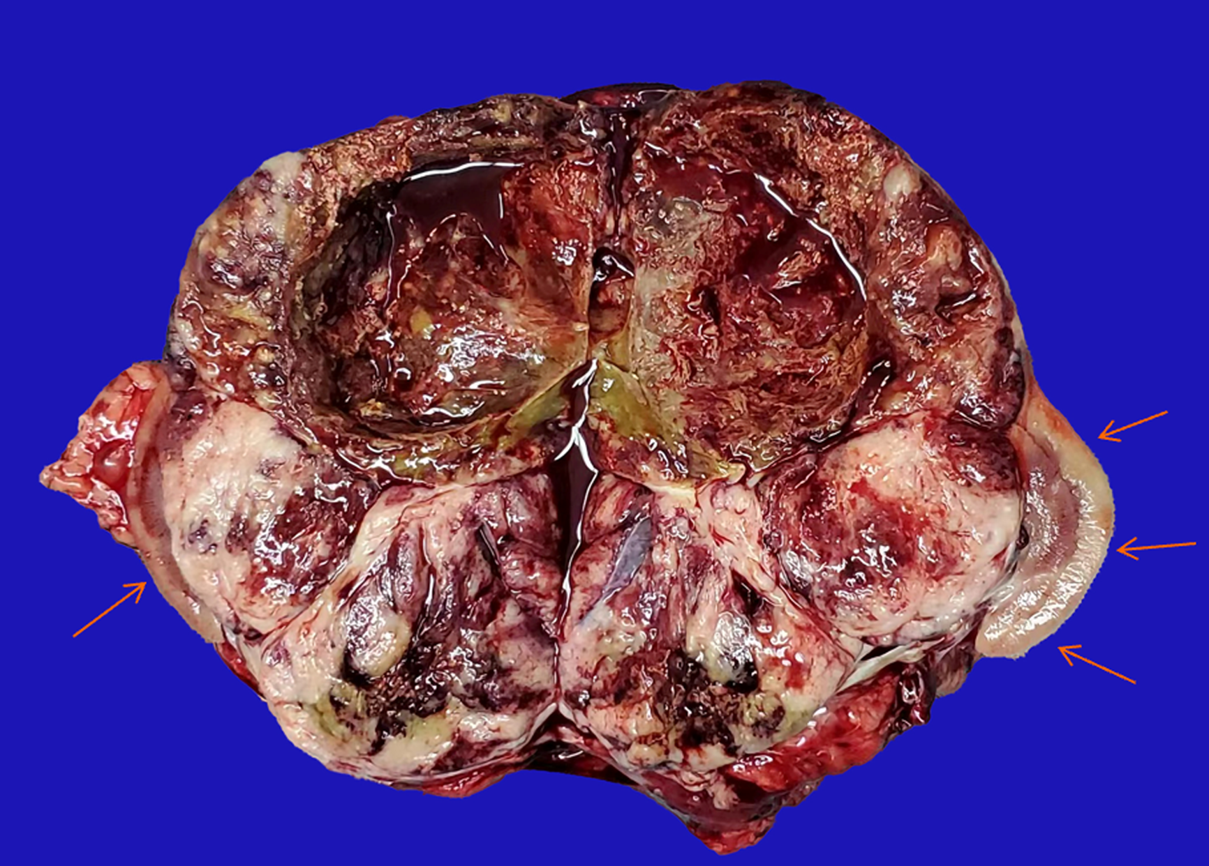
Figure 5 Gross specimen.
A huge tumor has invaded the entire kidney (coronal cut), leaving only a small portion of normal kidney tissue (Orange arrow). The internal cystic changes of the tumor were accompanied by bleeding and necrosis.
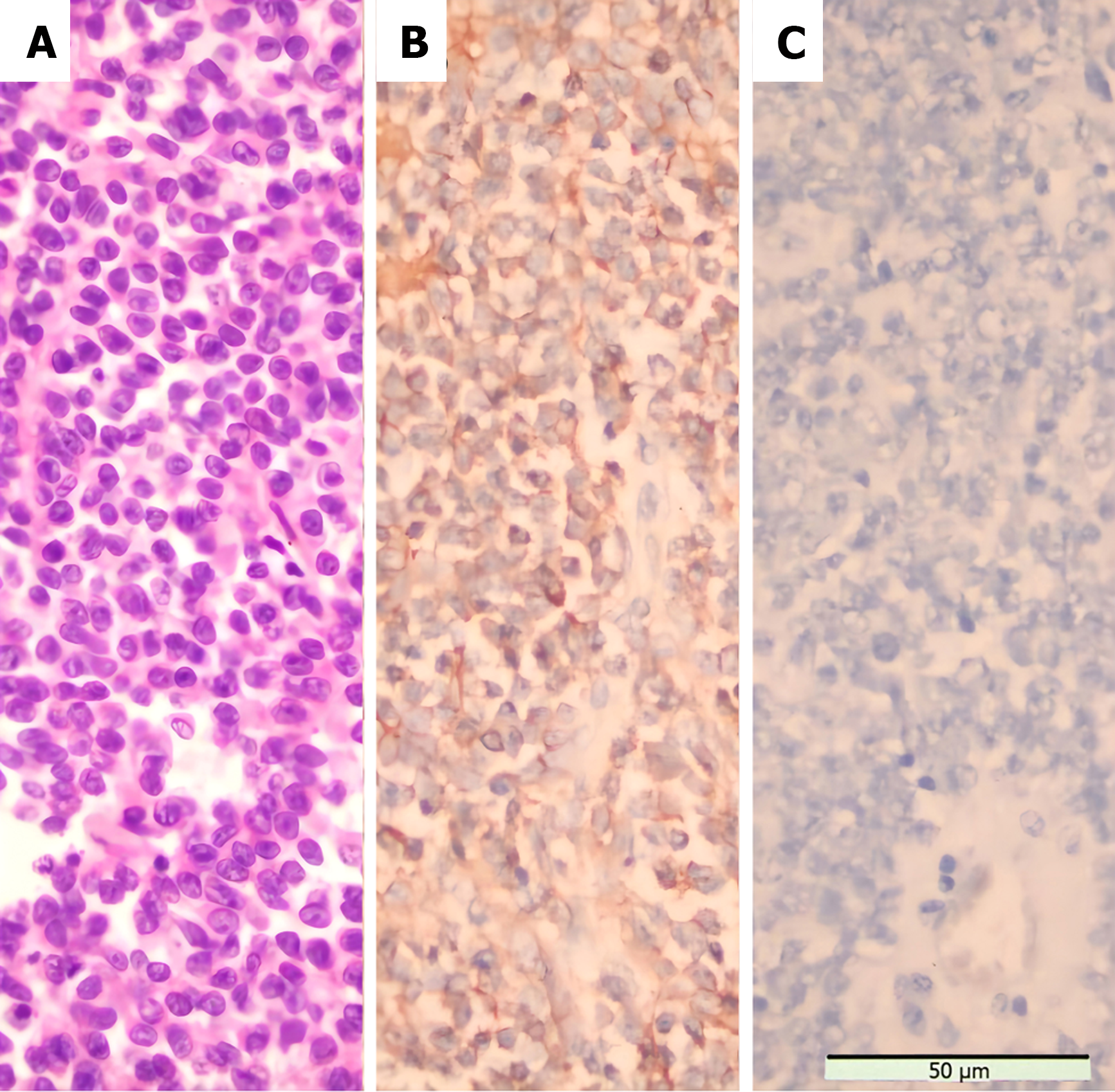
Figure 6 Morphological features and immunostaining results of the tumor.
A: Small, round tumor cells can be seen scattered throughout the visual field Staining method: HE staining (400 ×); B: Diffuse hyperstaining of tumor cell membrane for CD99 Staining method: Polymer method (400 ×); C: No positive reaction was observed for adrenocor ticotropic hormore staining method: Polymer method (400 ×).
- Citation: Dong GF, Hou YK, Ma Q, Ma SY, Wang YJ, Rexiati M, Wang WG. Cushing's syndrome caused by giant Ewing's sarcoma of the kidney: A case report and review of literature. World J Clin Cases 2024; 12(23): 5431-5440
- URL: https://www.wjgnet.com/2307-8960/full/v12/i23/5431.htm
- DOI: https://dx.doi.org/10.12998/wjcc.v12.i23.5431


