Copyright
©The Author(s) 2024.
World J Clin Cases. Aug 6, 2024; 12(22): 5229-5235
Published online Aug 6, 2024. doi: 10.12998/wjcc.v12.i22.5229
Published online Aug 6, 2024. doi: 10.12998/wjcc.v12.i22.5229
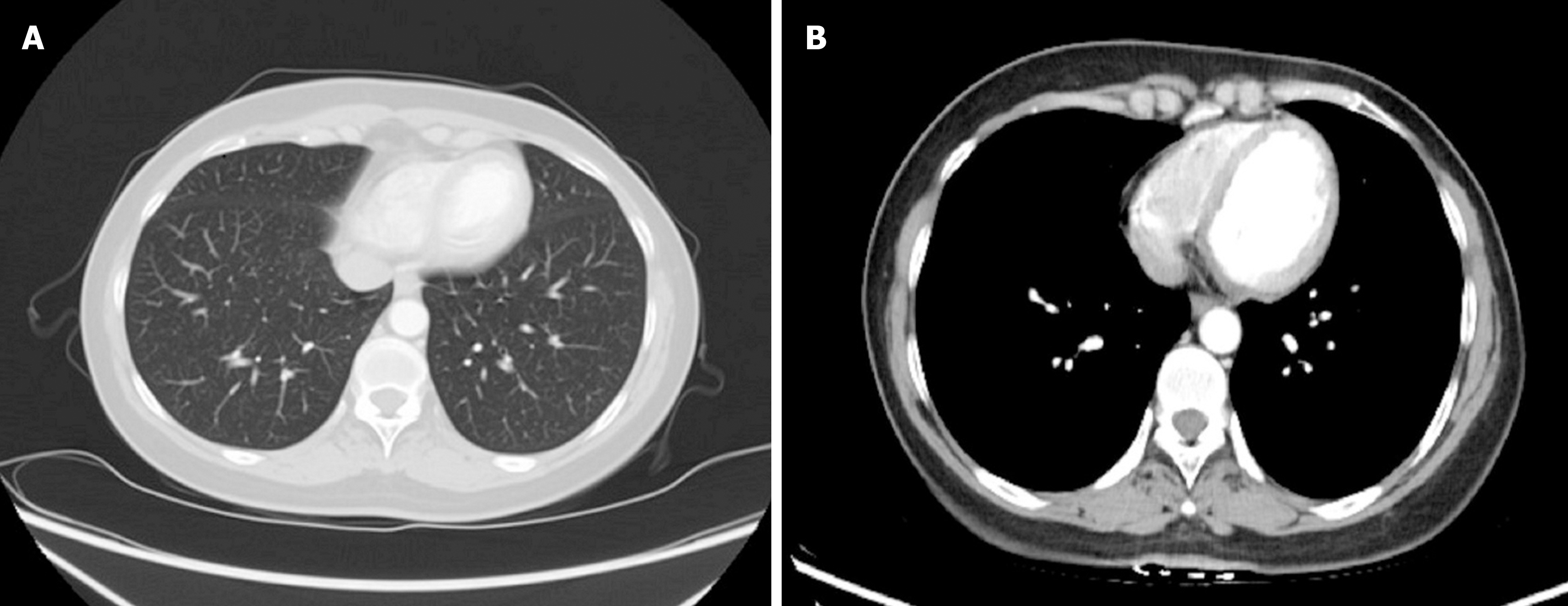
Figure 1 Chest and abdominal computed tomography findings.
A and B: Chest-abdominal plain computed tomography showed small nodules in both lungs. No other significant abnormalities were found.
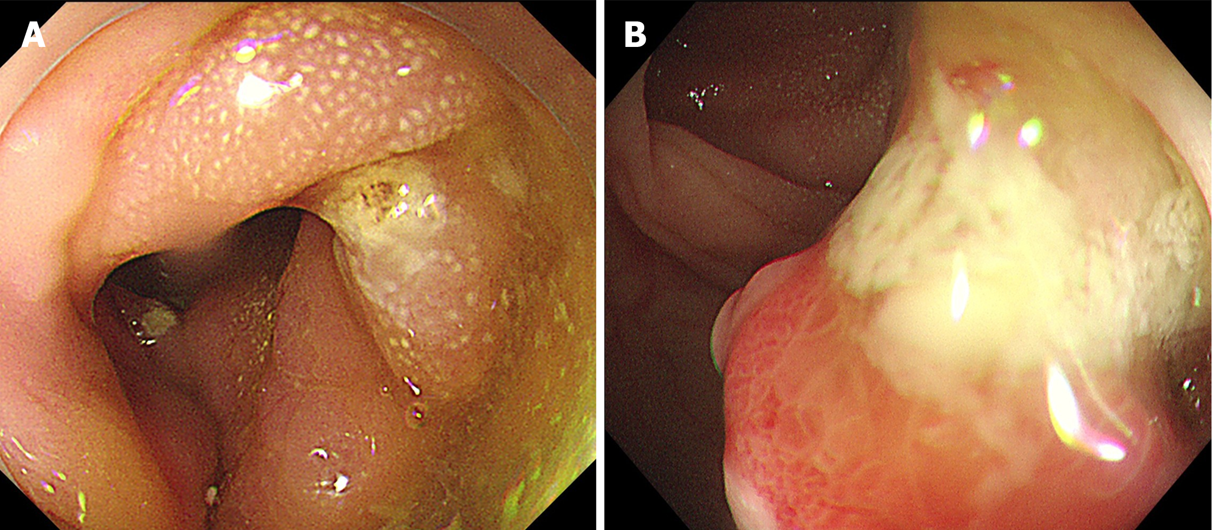
Figure 2 Endoscopic findings.
A and B: Colonoscopy image showing a pedunculated polyp in the transverse colon with a circumference of 5 cm.
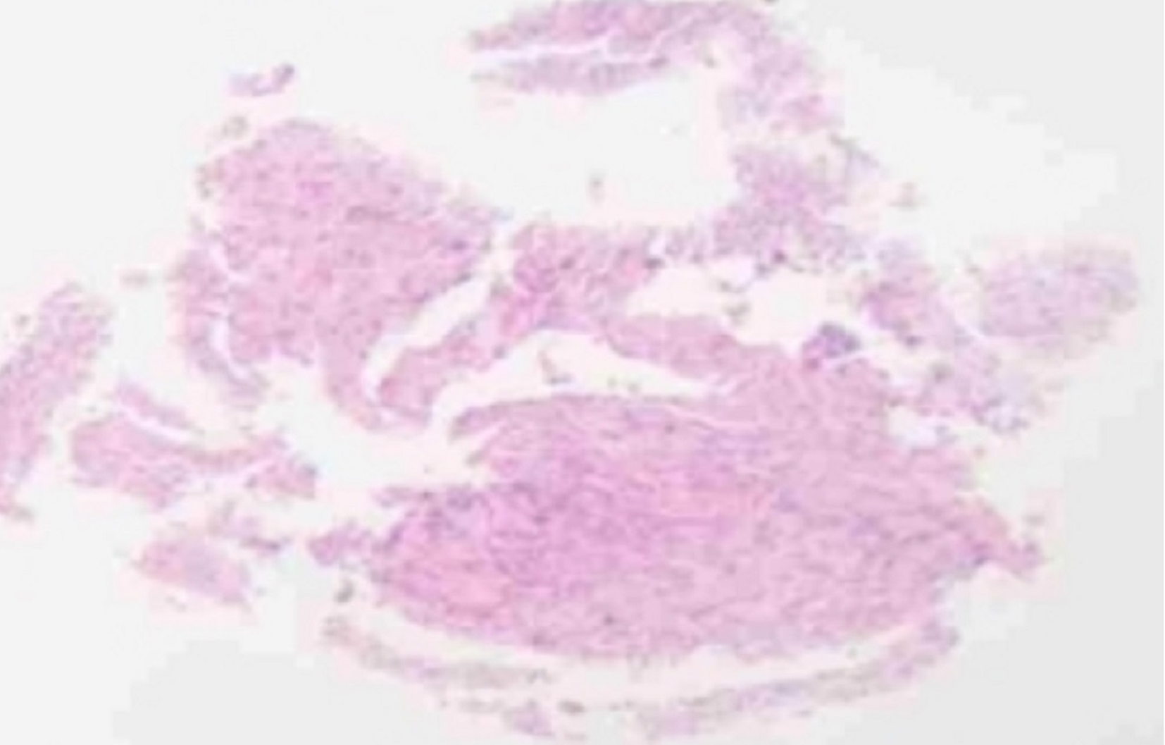
Figure 3
First pathology image showing heterogeneous nuclear cells in inflammatory granulation tissue.
Figure 4 Endoscopic submucosal dissection was performed for the lesion.
A and B: Submucosal injection of indigo carmine, epinephrine, and saline; C: Colonic polyp lesion.
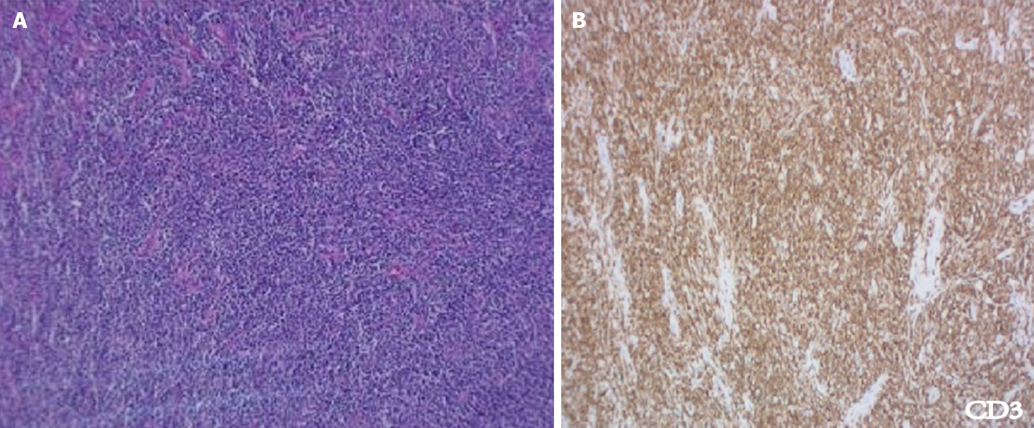
Figure 5 Pathology and immunohistochemistry.
A: Histopathology suggested highly active proliferation of T lymphocytes; B: CD3 immunohistochemical staining was positive.
- Citation: Sun YH, Lu SS, Fang Y, Xiong Z, Sun QY, Huang J. Rare primary colonic T cell lymphoma with curative resection by endoscopic submucosal dissection: A case report. World J Clin Cases 2024; 12(22): 5229-5235
- URL: https://www.wjgnet.com/2307-8960/full/v12/i22/5229.htm
- DOI: https://dx.doi.org/10.12998/wjcc.v12.i22.5229


