Copyright
©The Author(s) 2024.
World J Clin Cases. Aug 6, 2024; 12(22): 5217-5224
Published online Aug 6, 2024. doi: 10.12998/wjcc.v12.i22.5217
Published online Aug 6, 2024. doi: 10.12998/wjcc.v12.i22.5217
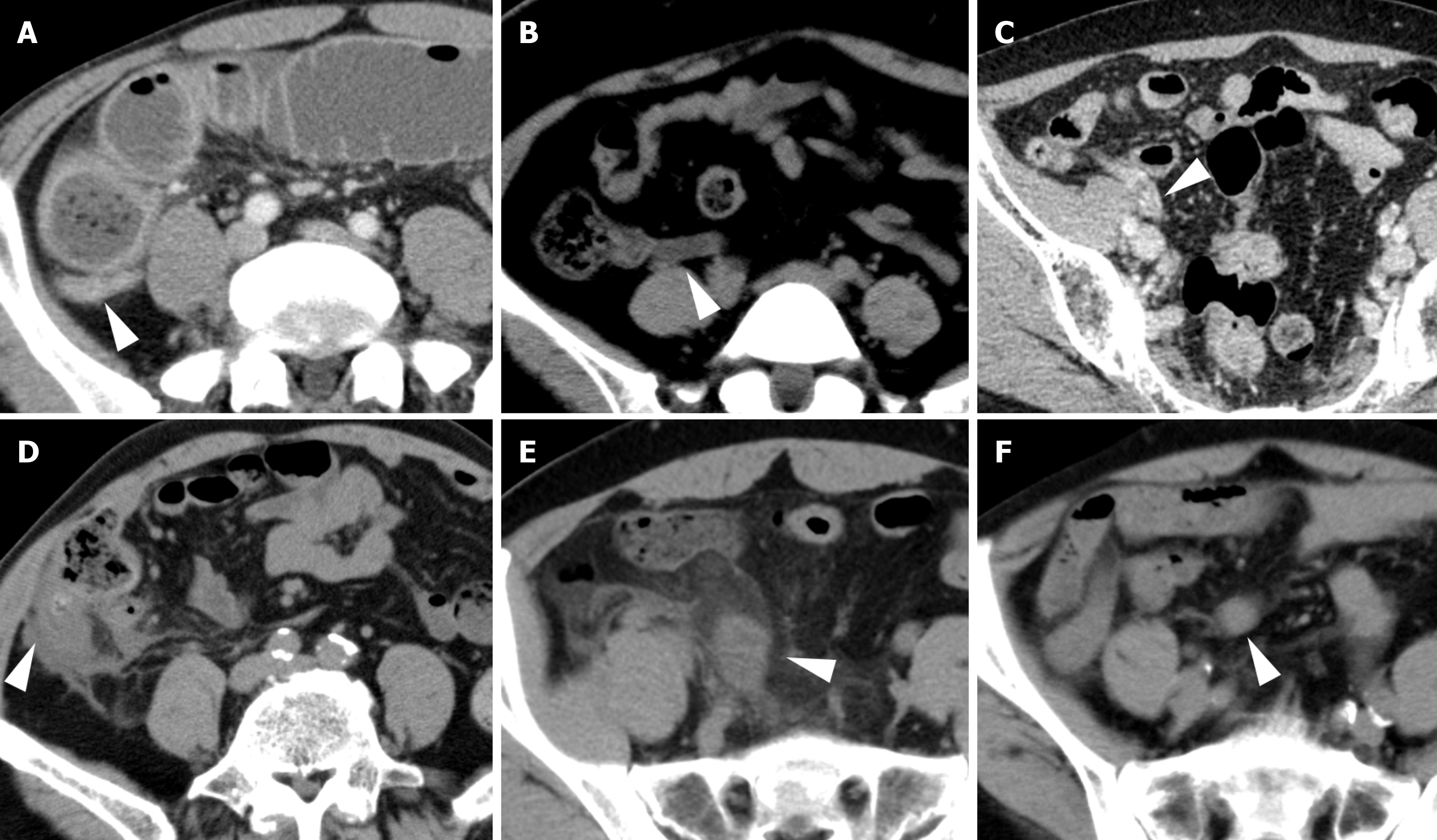
Figure 1 Abdominal computed tomography of the appendix in 6 patients.
Arrowheads indicate the appendix for each case. A: Case 1; B: Case 2; C: Case 3; D: Case 4; E: Case 5; F: Case 6.
Figure 2 Findings on colonoscopy.
A and B: In Case 1, colonoscopy shows edematous mucosa of the cecal wall (A), but no neoplastic changes are apparent at the cecum or appendiceal orifice (B); C: In Case 4, colonoscopy before additional ileocecal resection shows no neoplastic changes at the cecum or appendiceal orifice.
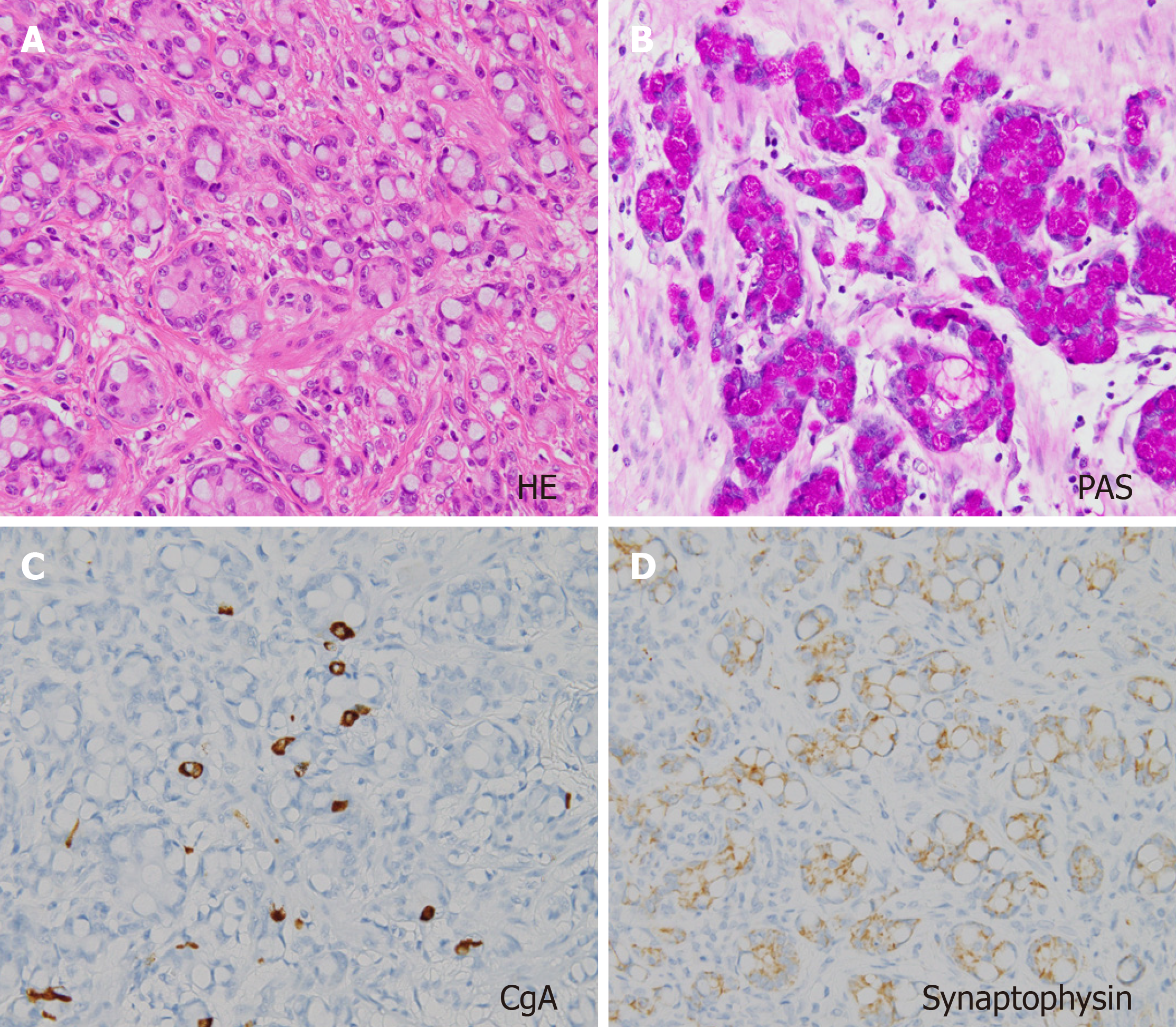
Figure 3 Representative pathological examination of goblet cell carcinoid in Case 1.
A: Hematoxylin and eosin (HE) staining shows a mixed pattern comprising large goblet cells mimicking signet-ring cells containing mucus; B: Periodic acid-Schiff (PAS) staining for mucin is positive; C and D: Immunohistochemical staining is positive for chromogranin A (CgA) (C) and synaptophysin (D).
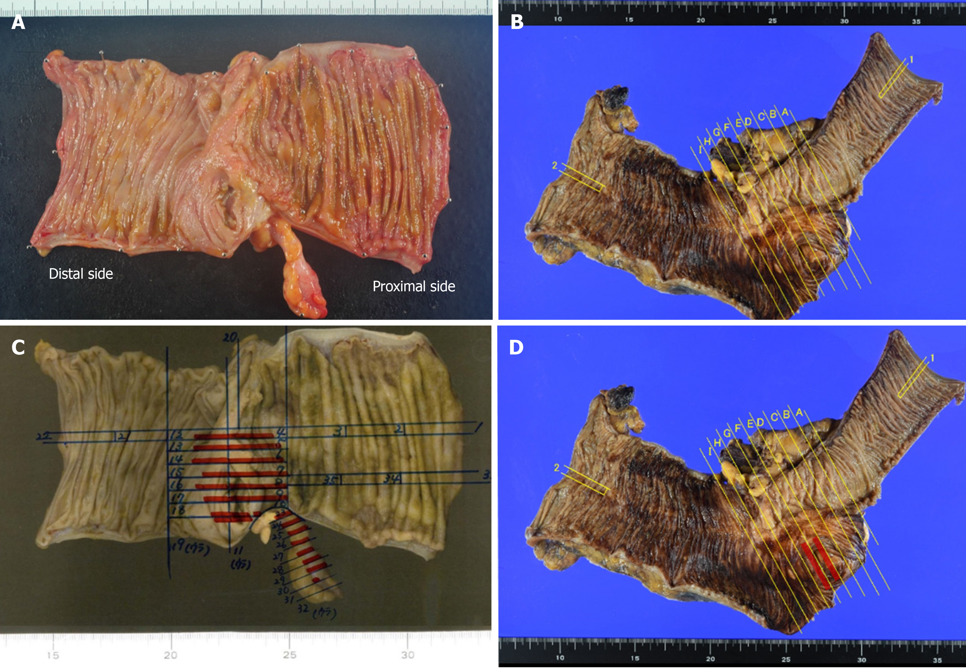
Figure 4 Resected specimen and tumor mapping.
A-C: Although no neoplastic changes are seen at the cecal surface or appendiceal orifice in Case 1 (A) or Case 4 (C), tumor cells have invaded widely into the cecum in Case 1 (B); D: Residual tumor cells are seen microscopically at the cecum around the appendiceal orifice in Case 4. Red lines show the existence of tumor cells.
- Citation: Toshima T, Inada R, Sakamoto S, Takeda E, Yoshioka T, Kumon K, Mimura N, Takata N, Tabuchi M, Oishi K, Sato T, Sui K, Okabayashi T, Ozaki K, Nakamura T, Shibuya Y, Matsumoto M, Iwata J. Goblet cell carcinoid of the appendix: Six case reports. World J Clin Cases 2024; 12(22): 5217-5224
- URL: https://www.wjgnet.com/2307-8960/full/v12/i22/5217.htm
- DOI: https://dx.doi.org/10.12998/wjcc.v12.i22.5217


