Copyright
©The Author(s) 2024.
World J Clin Cases. Jul 26, 2024; 12(21): 4827-4835
Published online Jul 26, 2024. doi: 10.12998/wjcc.v12.i21.4827
Published online Jul 26, 2024. doi: 10.12998/wjcc.v12.i21.4827
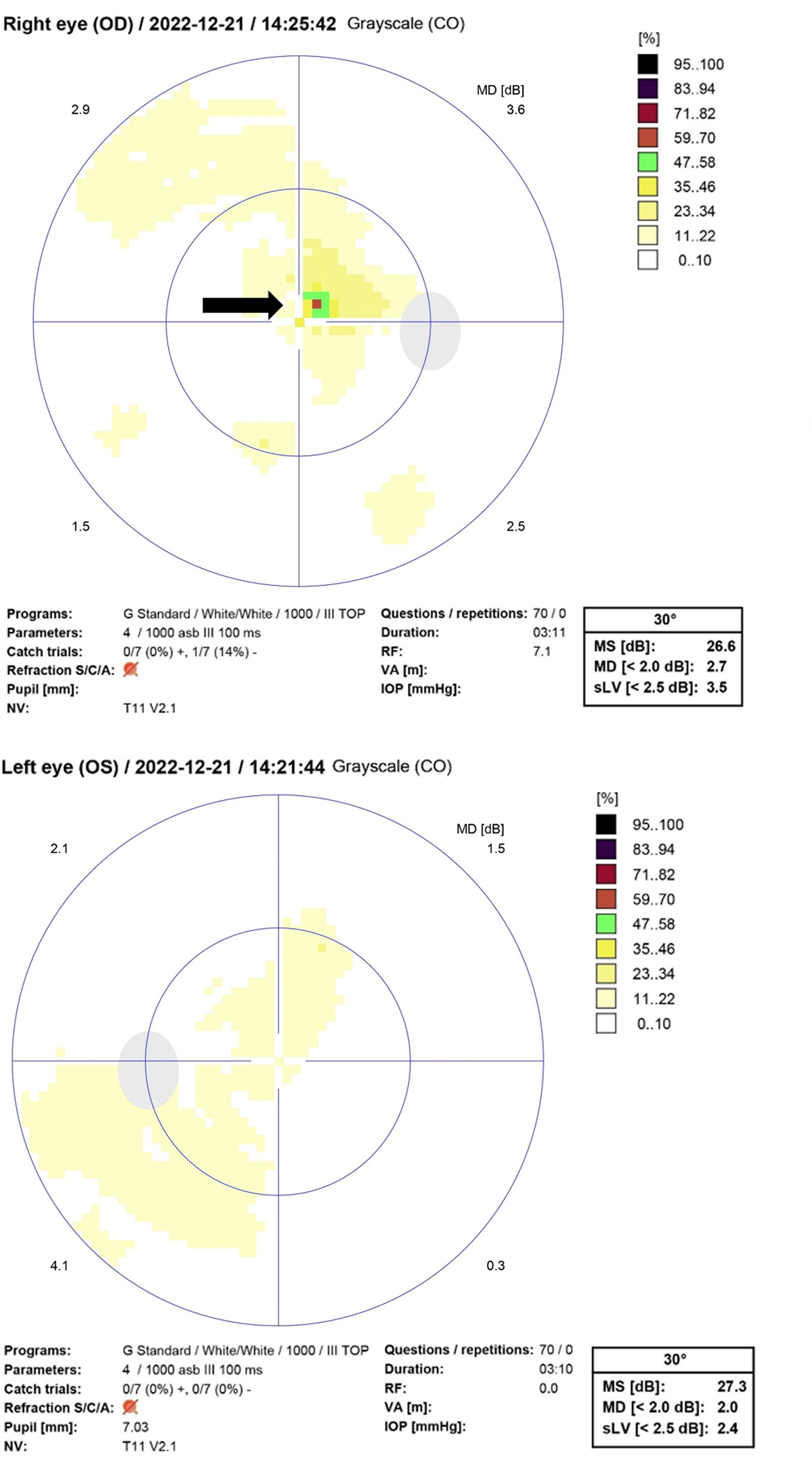
Figure 1
Central scotoma was shown in the perimetry of both eyes (black arrow).
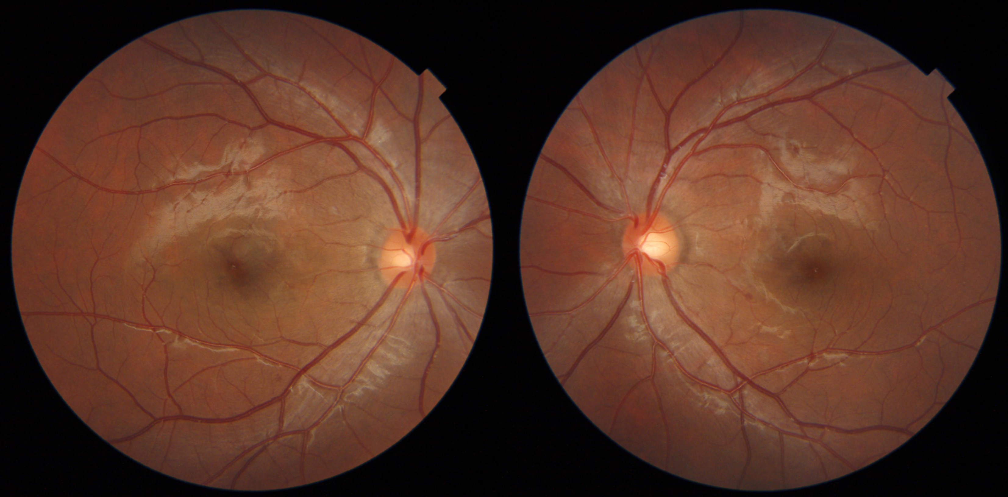
Figure 2
Images of bilateral normal fundus.
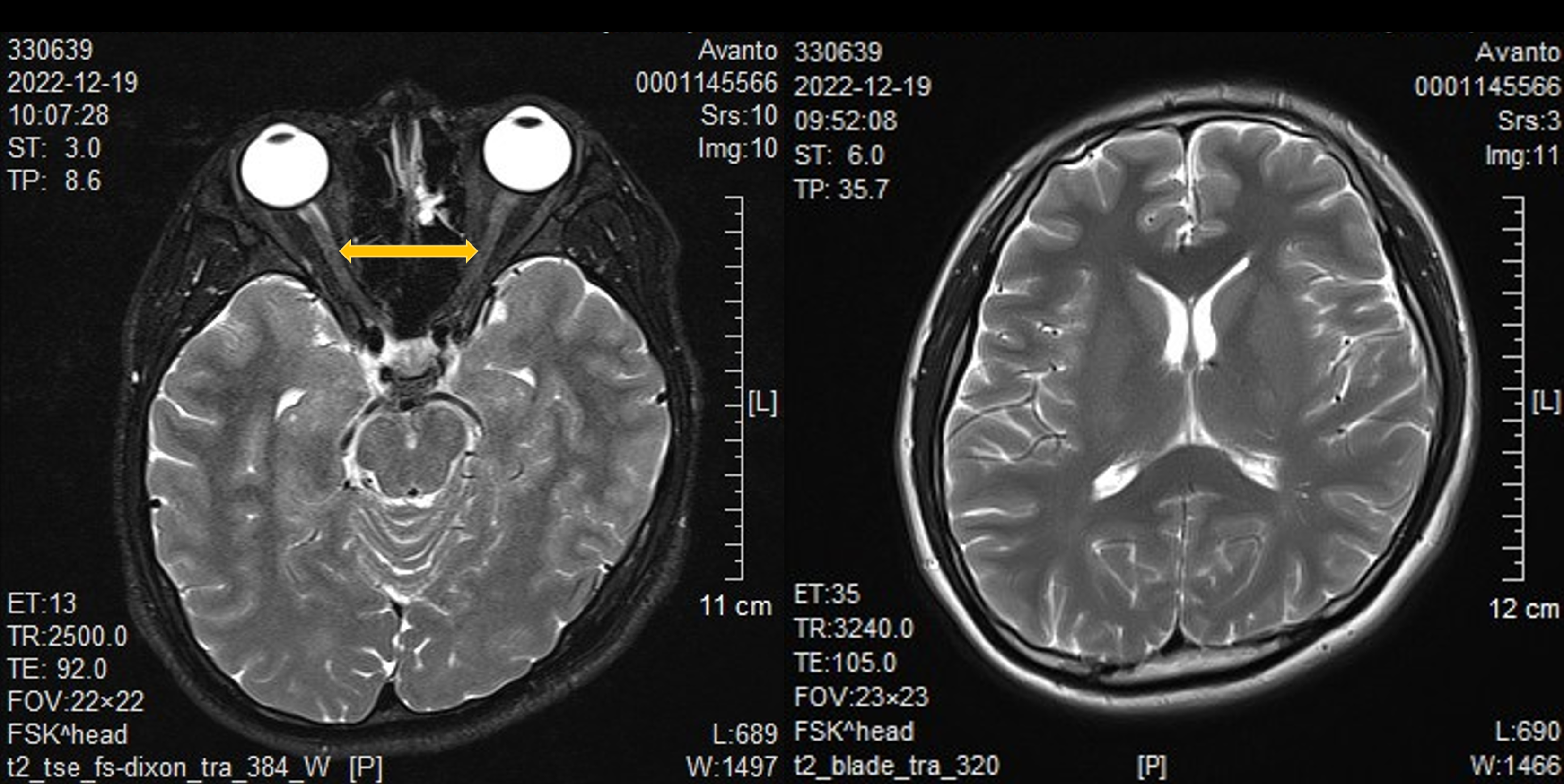
Figure 3
The orbital and brain magnetic resonance imaging results indicated an enlargement of the retrobulbar intraorbital segments of the optic nerve, which had a high T2 signal (yellow double arrow), and the brain tissue was in a healthy state.
- Citation: Li RR, Zhang BM, Rong SR, Li H, Shi PF, Wang YC. Fifteen acute retrobulbar optic neuritis associated with COVID-19: A case report and review of literature. World J Clin Cases 2024; 12(21): 4827-4835
- URL: https://www.wjgnet.com/2307-8960/full/v12/i21/4827.htm
- DOI: https://dx.doi.org/10.12998/wjcc.v12.i21.4827


