Copyright
©The Author(s) 2024.
World J Clin Cases. Jul 26, 2024; 12(21): 4820-4826
Published online Jul 26, 2024. doi: 10.12998/wjcc.v12.i21.4820
Published online Jul 26, 2024. doi: 10.12998/wjcc.v12.i21.4820
Figure 1 The physical examination we found that the patient had alopecia, onychodystrophy, and skin pigmentation.
A: The region of the head; B: Palms; C: Soles.
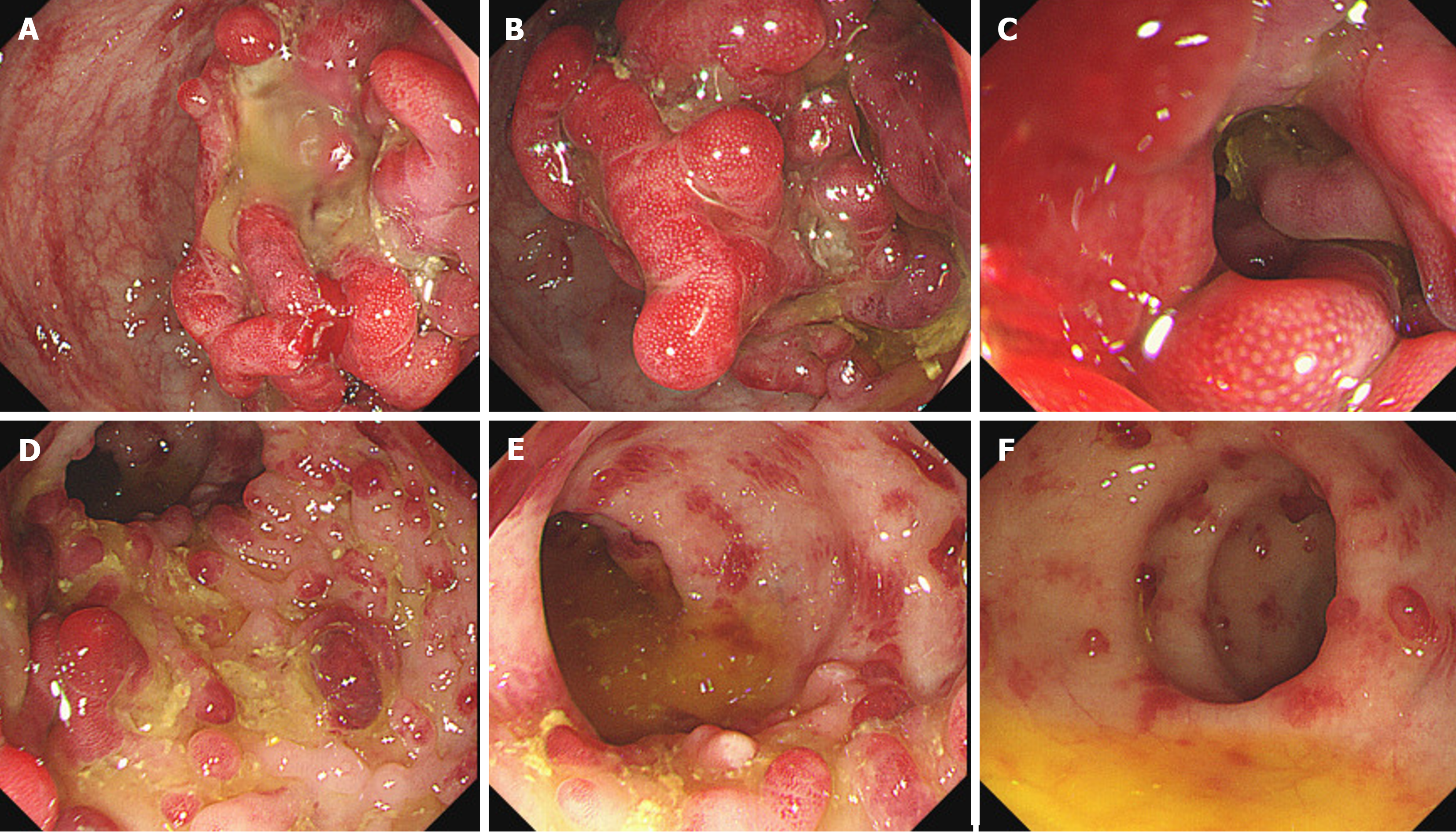
Figure 2 Enteroscopy.
A-C: The sigmoid colon: the mucosa of the whole colon was rough, and the left colon was dominated by red polypoid hyperplasia. The sigmoid colon from 15 cm to 20 cm at the entrance of the anus shows hyperplastic polyps growing in the circumferential cavity and the surface of the polyps is congested; D-F: Descending colon: there are mainly mucosal scars in the right colon, and red sessile polyps are diffused in the intestinal cavity.
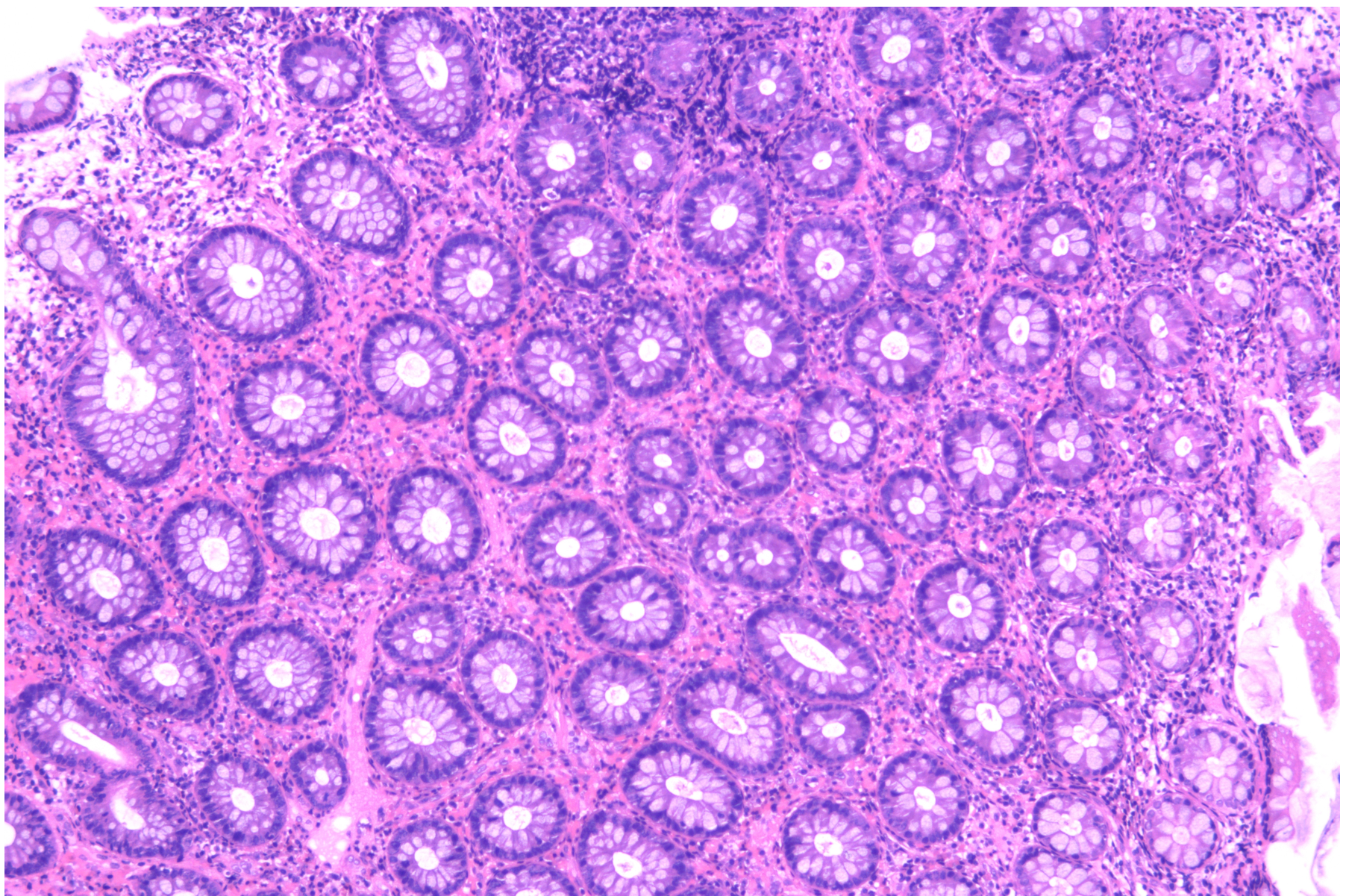
Figure 3 Histological photos of endoscopic biopsy specimens taken from the colon.
A biopsy of the sigmoid colon tissue showed chronic inflammation of the intestinal mucosa with sarcoid hyperplasia, glandular proliferative changes, and the formation of abscesses in individual lumens (hematoxylin-eosin staining, × 100).
- Citation: He ML, Zheng Y, Tian SX. Cronkhite-Canada syndrome complicated with pulmonary embolism: A case report. World J Clin Cases 2024; 12(21): 4820-4826
- URL: https://www.wjgnet.com/2307-8960/full/v12/i21/4820.htm
- DOI: https://dx.doi.org/10.12998/wjcc.v12.i21.4820


