Copyright
©The Author(s) 2024.
World J Clin Cases. Jul 16, 2024; 12(20): 4397-4404
Published online Jul 16, 2024. doi: 10.12998/wjcc.v12.i20.4397
Published online Jul 16, 2024. doi: 10.12998/wjcc.v12.i20.4397
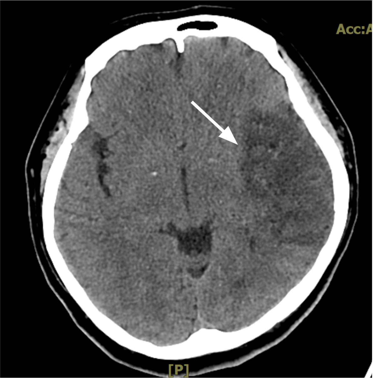
Figure 1 Coronary view of computer tomography of the brain.
Left cerebrum infarction was noted (arrow).
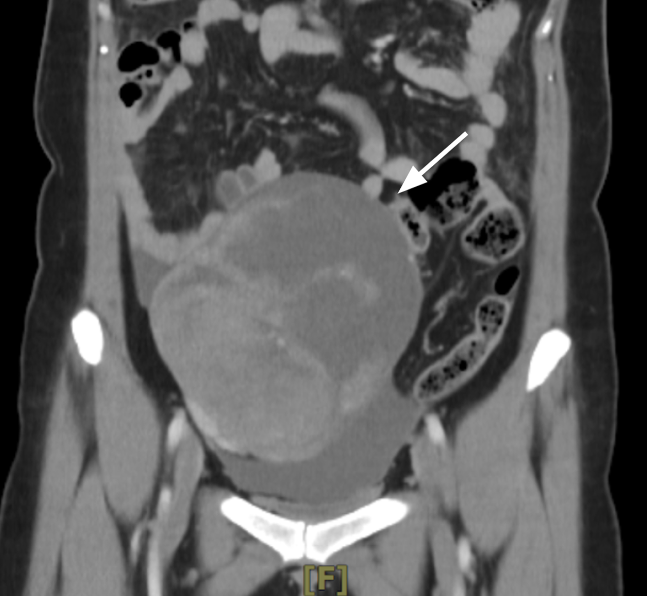
Figure 2 Coronary view of computer tomography of the abdominal cavity.
A 14.2 cm mixed cystic and solid components tumor was noted in the pelvic cavity (arrow).
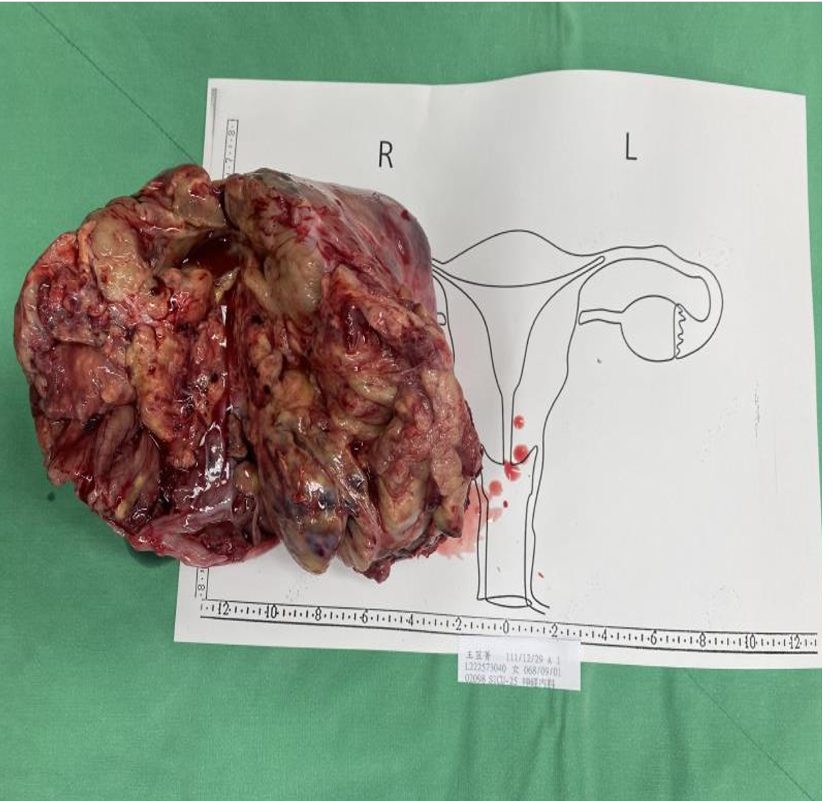
Figure 3 Gross picture of the right ovarian tumor.
A yellowish multi-lobulated ovarian tumor was noted.
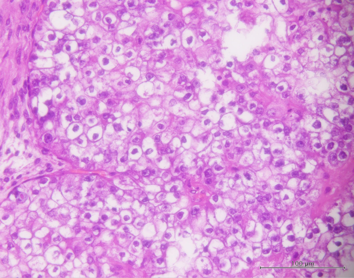
Figure 4 Pathological picture of the tumor.
Clear cell carcinoma of the ovary was noted. Clear cytoplasm and varied nuclei morphology (cubmoidal to large, polygonal nuclei) were noted. Scale bar = 100 μm.
- Citation: Siu WYS, Ding DC. Ischemic stroke with concomitant clear cell carcinoma of the ovary: A case report and review of literature. World J Clin Cases 2024; 12(20): 4397-4404
- URL: https://www.wjgnet.com/2307-8960/full/v12/i20/4397.htm
- DOI: https://dx.doi.org/10.12998/wjcc.v12.i20.4397


