Copyright
©The Author(s) 2024.
World J Clin Cases. Jul 16, 2024; 12(20): 4365-4371
Published online Jul 16, 2024. doi: 10.12998/wjcc.v12.i20.4365
Published online Jul 16, 2024. doi: 10.12998/wjcc.v12.i20.4365
Figure 1 Cranial magnetic resonance imaging of the patient.
A: T2WI high signal in the left maxillary sinus; B: symmetry of intracerebral structures, clear gray-white matter structures; C: the bilateral auditory nerves were well displayed, with no occupancy.
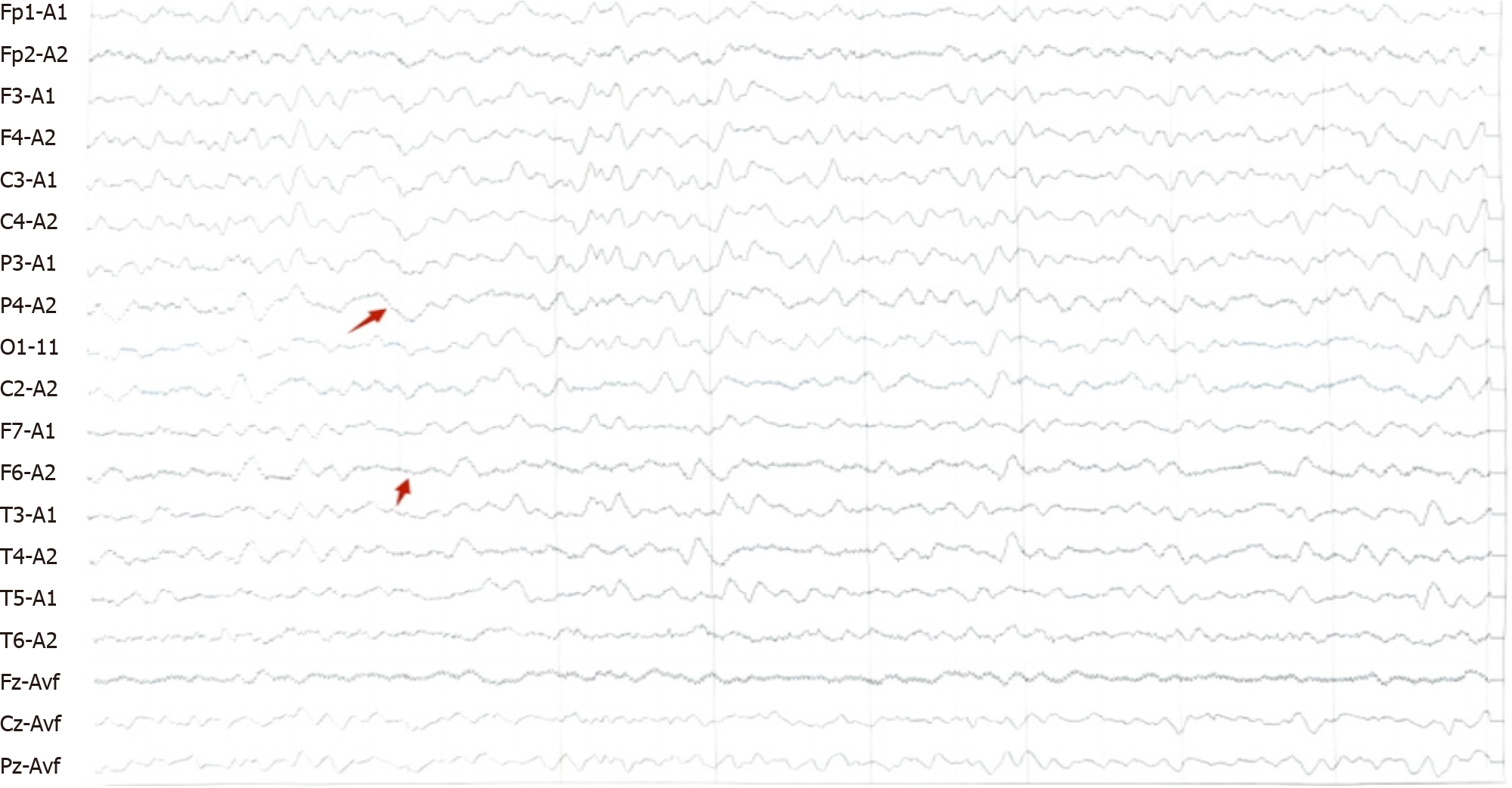
Figure 2 Long-range video electroencephalogram of the patient.
Increased sharp waves and slow waves in the left frontotemporal region indicated by the arrow.
- Citation: Chen HY, Wang J, Song DY, Wang B, Xu ZY, Wu Q, Wang ZL. Anti-contact protein-associated protein 2 antibody encephalitis in children: A case report. World J Clin Cases 2024; 12(20): 4365-4371
- URL: https://www.wjgnet.com/2307-8960/full/v12/i20/4365.htm
- DOI: https://dx.doi.org/10.12998/wjcc.v12.i20.4365


